The Acute Red Eye
A boards-focused triage of the acute red eye: separate the benign causes (viral/allergic conjunctivitis, subconjunctival hemorrhage) from the sight-threatening emergencies (angle-closure glaucoma, bacterial/HSV keratitis, anterior uveitis, scleritis, endophthalmitis) using vision, pain, pupil, injection pattern, and fluorescein — with the exact "next best step" the exam rewards.
Frame it: benign vs. sight-threatening
The acute red eye is a triage problem. Most cases are benign — viral or allergic conjunctivitis, subconjunctival hemorrhage, blepharitis, dry eye — and need only supportive care. But a short list of diagnoses can destroy vision within hours: bacterial/HSV keratitis, anterior uveitis, acute angle-closure glaucoma, scleritis, and endophthalmitis. Every red eye earns the same rapid sweep of discriminators: visual acuity, pain quality, the pupil, discharge vs. watery tearing, the pattern of injection (diffuse vs. ciliary/perilimbal flush), and the cornea on fluorescein.
Treat these as red flags demanding urgent ophthalmology: reduced visual acuity, deep/severe pain, true (consensual) photophobia, ciliary flush, a corneal opacity or fluorescein-staining defect, a fixed mid-dilated pupil, or contact lens wear. Two rules keep you out of trouble on exam day: don't dilate a red eye you haven't characterized (risks precipitating angle closure), and never start empiric topical steroids before excluding HSV and bacterial keratitis.
- Normal vision + itching/discharge + no true pain → benign (conjunctivitis, allergy, subconjunctival hemorrhage).
- ↓ Vision, deep pain, photophobia, or ciliary flush → sight-threatening until proven otherwise.
- Contact lens + painful red eye + corneal infiltrate = bacterial keratitis (Pseudomonas) until proven otherwise → scrape/culture, topical fluoroquinolone, never patch.
- Fixed mid-dilated pupil + steamy cornea + halos + headache/vomiting = acute angle-closure glaucoma → measure IOP now.
- Ciliary flush + photophobia + small/irregular pupil + cells & flare = anterior uveitis → topical steroid + cycloplegic; hunt HLA-B27 disease/sarcoid.
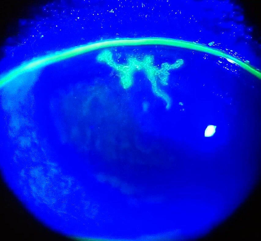
- Dendritic ulcer on fluorescein + ↓ corneal sensation = HSV epithelial keratitis → topical antiviral; steroids alone are contraindicated.
- Hyperacute copious purulent discharge in a sexually active adult = gonococcal conjunctivitis → Gram stain, IM ceftriaxone, co-treat chlamydia.
- Severe boring pain that wakes from sleep + violaceous sclera that won't blanch = scleritis → workup RA/GPA; systemic anti-inflammatory (not just topical).
- Painless, sharply bordered blood on the white of the eye = subconjunctival hemorrhage → reassurance.
One-look comparison
| Entity | Pain | Vision | Pupil | Cornea / injection clue | Next best step |
|---|---|---|---|---|---|
| Viral conjunctivitis | Gritty, no true pain | Normal | Normal | Diffuse redness, watery discharge, preauricular node | Supportive; strict hygiene (very contagious) |
| Allergic conjunctivitis | Itch | Normal | Normal | Bilateral, stringy mucus, chemosis | Topical antihistamine / mast-cell stabilizer |
| Subconjunctival hemorrhage | None | Normal | Normal | Focal, sharply bordered blood | Reassure; check BP / anticoagulation if recurrent |
| Bacterial keratitis | Yes | ↓ | Normal | Infiltrate/ulcer ± hypopyon, contact lens | Scrape/culture + topical fluoroquinolone; don't patch |
| Anterior uveitis | Deep, photophobia | Normal/↓ | Small, irregular | Ciliary flush, cells & flare | Topical steroid + cycloplegic; HLA-B27 / sarcoid workup |
| Acute angle-closure glaucoma | Severe + HA/nausea | ↓, halos | Mid-dilated, fixed | Steamy cornea, hard globe | Measure IOP, lower it, laser iridotomy |
| Scleritis | Severe, boring, wakes from sleep | Normal/↓ | Normal | Violaceous, no blanch w/ phenylephrine; RA/GPA | Systemic NSAID/steroid; autoimmune workup |
| Episcleritis | Mild/none | Normal | Normal | Sectoral, blanches w/ phenylephrine | Reassure ± NSAID; self-limited |
A 62-year-old woman develops sudden severe right eye pain, blurred vision with halos around lights, frontal headache, nausea, and vomiting shortly after sitting in a dark movie theater. The conjunctiva is diffusely red, the cornea is hazy/steamy, the pupil is mid-dilated and fixed, and the globe feels rock-hard.
Buzzwords: halos around lights, mid-dilated fixed pupil, steamy cornea, nausea/vomiting (can masquerade as a GI or neuro emergency), precipitated by dim light, mydriatics, or anticholinergics.
Next best step: Measure the intraocular pressure (tonometry) — a markedly elevated IOP confirms it. Then lower the IOP urgently: topical timolol + apraclonidine (or brimonidine) + pilocarpine plus oral/IV acetazolamide (± IV mannitol), and call ophthalmology for definitive laser peripheral iridotomy. Note that pilocarpine is often ineffective until the pressure falls (very high IOP causes iris-sphincter ischemia), so it's typically added once the other agents lower the pressure. Do not dilate the pupil.
(A) Bacterial keratitis — A 20-year-old who sleeps in soft contact lenses has 2 days of a painful red eye, photophobia, tearing, and blurred vision. Slit lamp shows a round white corneal infiltrate that stains with fluorescein, with a small hypopyon. Think Pseudomonas. Next best step: corneal scraping for Gram stain and culture, then topical fluoroquinolone (or fortified antibiotics); stop lens wear; do not patch and do not give topical steroids.
(B) HSV keratitis — A patient with recurrent unilateral painful red eyes has a branching dendritic ulcer with terminal bulbs on fluorescein and decreased corneal sensation. Next best step: topical (or oral) antiviral (e.g., ganciclovir gel / trifluridine, or oral acyclovir/valacyclovir); withhold topical steroids in epithelial disease — they can drive a geographic ulcer.
(A) Anterior uveitis — A 28-year-old man with chronic inflammatory low-back stiffness has a painful, photophobic red eye. Exam shows ciliary (perilimbal) flush, a small/irregular pupil, and cells and flare in the anterior chamber. Next best step: slit-lamp confirmation, then topical corticosteroid + cycloplegic (relieves pain, prevents posterior synechiae), plus a systemic workup — HLA-B27 spondyloarthropathies, sarcoidosis, and infection (syphilis, TB, HSV/VZV).
(B) Gonococcal conjunctivitis — A sexually active adult has hyperacute (< 24 h), copious purulent discharge, marked lid swelling, and chemosis; Gram stain shows gram-negative intracellular diplococci. It can perforate the cornea — an emergency. Next best step: single-dose IM ceftriaxone + saline irrigation, and co-treat for chlamydia (doxycycline unless excluded).

PAIR — the HLA-B27 seronegative spondyloarthropathies that cause anterior uveitis:
- Psoriatic arthritis
- Ankylosing spondylitis
- Inflammatory bowel disease (enteropathic arthritis)
- Reactive arthritis
Reactive arthritis triad — "can't see, can't pee, can't climb a tree" → conjunctivitis (± uveitis) + urethritis + arthritis (post-GU/GI infection).
- Suspected angle-closure → tonometry first; avoid mydriatics.
- Contact lens + infiltrate → scrape/culture + topical fluoroquinolone; never patch.
- Photophobia + ciliary flush → slit lamp for cells/flare → steroid + cycloplegic + systemic workup.
- Dendrite on fluorescein → antiviral, not steroid.
- Hyperacute purulent discharge → Gram stain + IM ceftriaxone + chlamydia coverage.
- Post-op or post-intravitreal-injection painful red eye with hypopyon → suspect endophthalmitis → emergent vitreous tap + intravitreal antibiotics.
- Trauma → rule out open globe (positive Seidel test), hyphema, retained foreign body → rigid shield, no pressure, CT orbit, update tetanus.
- Do-not-miss reflex: undiagnosed red eye → no empiric steroids, no dilation, refer on any red flag.
Practice Ophthalmology now
Board-style questions, spaced-repetition flashcards, and a Socratic AI tutor — free to start.