Tachyarrhythmias: SVT, Atrial Fibrillation & VT
A board-focused walkthrough of tachyarrhythmias organized by the exam's own logic — narrow vs wide, regular vs irregular, stability first — covering SVT, atrial fibrillation/flutter, and VT with classic vignette buzzwords, ECG findings, and next-best-step management.
The Board Framework: Narrow vs Wide, Regular vs Irregular
Tachyarrhythmia = HR >100 bpm. On the boards, read every rhythm strip with two questions:
- QRS width — narrow (<120 ms → supraventricular) vs wide (≥120 ms → ventricular until proven otherwise).
- Regularity — regular vs irregular.
This 2×2 sorts nearly everything:
- Narrow + regular: sinus tach, AVNRT, AVRT, atrial flutter, atrial tachycardia
- Narrow + irregular: atrial fibrillation, MAT, flutter with variable block
- Wide + regular: monomorphic VT (most common), SVT with aberrancy
- Wide + irregular: AFib with aberrancy/pre-excitation (WPW), polymorphic VT/torsades
Before the diagnosis, decide stability. Hypotension, altered mentation, ischemic chest pain, or pulmonary edema = unstable → immediate synchronized cardioversion (pulseless VT/VF → unsynchronized defibrillation). Stability, not the exact rhythm, drives the first move.
- Unstable + any tachyarrhythmia → synchronized cardioversion now (pulseless VT/VF → defibrillation)
- Stable narrow-complex regular SVT: vagal maneuvers → IV adenosine 6 mg rapid push with saline flush, then 12 mg if needed
- Adenosine both terminates AVNRT/AVRT and unmasks atrial flutter/atrial tach (transient AV block reveals flutter waves)
- New AFib: rate control (β-blocker or non-dihydropyridine CCB) is first-line for most; anticoagulate by CHA₂DS₂-VASc. Avoid non-DHP CCBs (diltiazem/verapamil) in HFrEF or decompensated HF — negative inotropes; use a β-blocker or digoxin.
- Pre-excited AFib (WPW — wide, irregular): avoid AV-nodal blockers (adenosine, β-blockers, CCB, digoxin) → procainamide or cardioversion
- Wide-complex tachycardia + prior MI / structural heart disease = VT until proven otherwise
- Cardioverting AFib present >48 h (or unknown duration) requires 3 weeks anticoagulation OR TEE to exclude LA thrombus first
- Torsades → IV magnesium sulfate
Vignette: A 24-year-old woman has sudden-onset palpitations and lightheadedness. BP 118/76, HR 185, regular. ECG: narrow-complex regular tachycardia with no discernible P waves (buried in or just after the QRS as a pseudo-R′).
- Diagnosis: AVNRT — the most common paroxysmal SVT; classically young, healthy, with abrupt on/off palpitations.
- Next step (stable): vagal maneuvers (modified Valsalva, carotid sinus massage) → if unsuccessful, IV adenosine 6 mg rapid push, then 12 mg.
- If unstable (hypotension, syncope): synchronized cardioversion.
- Recurrent/definitive: catheter ablation of the AV-nodal slow pathway is curative.
Tachyarrhythmias at a Glance
| Arrhythmia | ECG hallmark | Classic clue | First-line Rx |
|---|---|---|---|
| AVNRT / AVRT (PSVT) | Narrow, regular, 150–250, no visible P | Young, abrupt palpitations | Vagal → adenosine |
| Atrial flutter | Sawtooth waves, atrial ~300, often 2:1 | Regular rate ~150 | Rate control, anticoag, CTI ablation (curative) |
| Atrial fibrillation | Irregularly irregular, no P waves | "Irregularly irregular" | Rate control + anticoag (CHA₂DS₂-VASc) |
| MAT | ≥3 distinct P-wave morphologies | COPD / hypoxia | Treat cause; verapamil |
| Monomorphic VT | Wide, regular; AV dissociation, fusion/capture beats | Prior MI / structural HD | Stable: amiodarone; unstable: cardiovert |
| Torsades (PMVT) | Polymorphic, twisting axis, long QT | QT-prolonging drugs, ↓K / ↓Mg | IV magnesium |

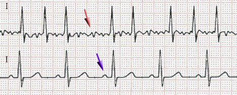
Vignette: A 72-year-old man with hypertension and diabetes has several hours of palpitations and mild dyspnea. BP 138/84, HR 148, irregularly irregular. ECG: no P waves, chaotic fibrillatory baseline, narrow QRS.
- Diagnosis: Atrial fibrillation with rapid ventricular response.
- Next step (stable): rate control — IV metoprolol or diltiazem to relieve symptoms.
- Anticoagulation: CHA₂DS₂-VASc = HTN (1) + DM (1) + age 65–74 (1) = 3 → start a DOAC (preferred over warfarin unless mechanical valve or moderate–severe mitral stenosis).
- Cardioversion caution: symptom onset ≠ confirmed rhythm onset, so when duration is ≥48 h or uncertain, do not electively cardiovert without 3 weeks anticoagulation or a TEE first.
- C — Congestive heart failure / LV dysfunction … 1
- H — Hypertension … 1
- A₂ — Age ≥75 … 2
- D — Diabetes … 1
- S₂ — prior Stroke / TIA / thromboembolism … 2
- V — Vascular disease (prior MI, PAD, aortic plaque) … 1
- A — Age 65–74 … 1
- Sc — Sex category (female) … 1
Anticoagulate: men ≥2 / women ≥3 → oral anticoagulant recommended, DOAC preferred. Men =1 / women =2 → intermediate; anticoagulation is reasonable (shared decision). Men 0 / women 1 (sex-only) → none. Pair with HAS-BLED to gauge bleeding risk — a high score flags modifiable bleeding factors and prompts caution but does not by itself contraindicate anticoagulation.
Vignette: A 68-year-old man with a prior anterior MI reports palpitations and chest pressure. BP 86/58, HR 172. ECG: wide-complex regular tachycardia with AV dissociation, fusion and capture beats.
- Diagnosis: Monomorphic ventricular tachycardia. With prior MI / structural heart disease, wide + regular = VT until proven otherwise — never reflexively call it "SVT with aberrancy."
- Next step: he is unstable (hypotension, ischemia) but has a pulse → synchronized cardioversion (pulseless → defibrillation).
- If stable: IV amiodarone (alternatives: procainamide, lidocaine); then treat underlying ischemia/cardiomyopathy and evaluate for an ICD.
- Trap: never give AV-nodal blockers (e.g., verapamil) to undifferentiated wide-complex tachycardia — can precipitate collapse.
- Pre-excited AFib (WPW): wide, irregular, rates can exceed 250. AV-nodal blockers are dangerous (adenosine, β-blockers, non-DHP CCB, digoxin) — they force conduction down the accessory pathway → risk of VF. Use procainamide or ibutilide, or cardiovert.
- Torsades de pointes: polymorphic VT on a long QT — give IV magnesium sulfate (even if the level is normal), correct K⁺/Mg²⁺, stop offending drugs; overdrive pacing or isoproterenol for pause-dependent forms.
- MAT: ≥3 P-wave morphologies, tied to COPD / hypoxia — treat the lung disease; verapamil if rate control needed; avoid nonselective β-blockers.
- Atrial flutter: same anticoagulation rules as AFib; cavotricuspid isthmus ablation is highly curative.
- Regular narrow SVT that won't break with adenosine: reconsider atrial flutter or atrial tachycardia.

Practice Cardiology now
Board-style questions, spaced-repetition flashcards, and a Socratic AI tutor — free to start.