T-Cell Development & Activation
A Step 1 high-yield walkthrough of how T cells are educated in the thymus (positive/negative selection, MHC restriction, AIRE) and activated in the periphery (two/three-signal model, checkpoints, Th subsets), each step mapped to the classic immunodeficiency or autoimmune vignette its defect produces.
The Two Jobs: Get Educated, Then Get Activated
T lymphocytes arise from bone-marrow hematopoietic progenitors but mature in the thymus ("T" = thymus). A developing thymocyte must survive two make-or-break checkpoints before release into the circulation.
- In the cortex, immature CD4+CD8+ double-positive cells undergo positive selection: a TCR that can bind self-MHC survives; one that cannot dies by neglect. This is what enforces MHC restriction.
- In the medulla, negative selection deletes clones whose TCR binds self-peptide too strongly — this is central tolerance. The AIRE gene lets medullary epithelial cells display tissue-specific self-antigens so autoreactive clones can be caught and killed.
Cells then commit to a single lineage — CD8 (reads MHC I) or CD4 (reads MHC II) — and exit as naive single-positive T cells. In the periphery, a naive T cell needs two signals (antigen + costimulation) to activate; antigen alone → anergy. Everything the boards test here is a variation on "which step broke, and what infection or autoimmune pattern results."
- Site: made in bone marrow, matured in the thymus (journey runs cortex → medulla).
- Thymocyte stages: double-negative (CD4−CD8−) → double-positive (CD4+CD8+) → single-positive (CD4+ or CD8+).
- TCR gene rearrangement uses RAG-1/RAG-2 (with TdT); failure blocks both TCR and BCR assembly → SCID.
- Positive selection = cortex: keep TCRs that bind self-MHC → sets MHC restriction; failure = death by neglect.
- Negative selection = medulla: delete TCRs with high affinity for self-peptide → central tolerance; depends on AIRE-displayed self-antigens.
- Lineage rule: TCR + MHC I → CD8 T cell; TCR + MHC II → CD4 T cell.
- Thymus fails to form → DiGeorge (del 22q11.2): low T cells, hypocalcemia, conotruncal cardiac defects, recurrent viral/fungal infection.
Thymic Education — Step by Step
| Checkpoint | Location | What is tested | Molecules / genes | Failure |
|---|---|---|---|---|
| TCR rearrangement | Cortex | Build a functional TCR | RAG-1/RAG-2, TdT | No TCR → SCID |
| Positive selection | Cortex | TCR binds self-MHC (any) | Cortical epithelial MHC I/II | Death by neglect |
| Lineage commitment | Cortex → medulla | Which MHC class is read | CD4↔MHC II, CD8↔MHC I | — |
| Negative selection | Medulla | TCR not strongly self-reactive | AIRE-driven self-antigens | AIRE loss → APECED (APS-1) |
Activation — The Three-Signal Model
A naive T cell is primed by a professional antigen-presenting cell — chiefly a dendritic cell (macrophages and B cells also present antigen, but dendritic cells are the key initiators of naïve-T-cell priming) — delivering up to three signals:
- Signal 1 — antigen: the TCR–CD3 complex engages peptide on MHC; the coreceptor stabilizes it — CD4 with MHC II, CD8 with MHC I.
- Signal 2 — costimulation: B7 (CD80/CD86) on the APC binds CD28 on the T cell. Signal 1 without signal 2 → anergy (peripheral tolerance).
- Signal 3 — cytokines: polarize the CD4 helper into a subset (IL-12 → Th1, IL-4 → Th2).
Downstream, TCR engagement raises intracellular Ca²⁺, activating calcineurin → NFAT (plus NF-κB and AP-1) to drive IL-2 and clonal expansion — the step cyclosporine/tacrolimus block. CTLA-4 (CD152) and PD-1 are inhibitory receptors that brake this response and are the targets of checkpoint-inhibitor cancer drugs (ipilimumab = anti-CTLA-4; nivolumab/pembrolizumab = anti-PD-1). Activated CD4 cells upregulate CD40L (CD154) to license B cells and macrophages.
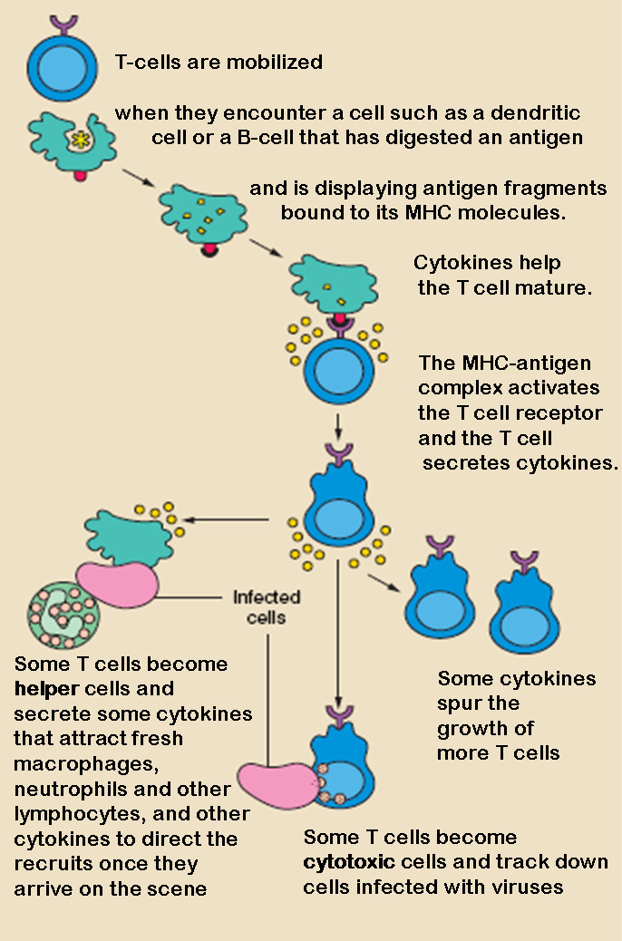
- Two-signal rule: Signal 1 = TCR–peptide/MHC; Signal 2 = B7–CD28. No signal 2 → anergy.
- Coreceptors: CD4 → MHC II, CD8 → MHC I (Rule of 8).
- CD3 transduces the TCR signal; ZAP-70 is the key kinase — deficiency = SCID variant with absent CD8 and nonfunctional CD4.
- Calcineurin → NFAT → IL-2; blocked by cyclosporine / tacrolimus.
- CTLA-4 and PD-1 = inhibitory checkpoints → cancer-immunotherapy targets.
- CD40L (T cell) – CD40 (B cell): required for Ig class switching; defect → Hyper-IgM syndrome.
- Superantigens (TSST-1, staph/strep enterotoxins) bridge MHC II to the TCR Vβ region outside the peptide groove → massive polyclonal T-cell activation → cytokine storm/toxic shock.
CD4 Helper Subsets — Comparison
| Subset | Induced by | Master TF | Key cytokines | Main job | Clinical link |
|---|---|---|---|---|---|
| Th1 | IL-12 | T-bet | IFN-γ, IL-2 | Activate macrophages & CD8 cells vs intracellular microbes | IL-12/IFN-γ-axis defect → disseminated mycobacteria/Salmonella |
| Th2 | IL-4 | GATA3 | IL-4, IL-5, IL-13 | Help B cells (IgE), recruit eosinophils vs helminths | Drives allergy/asthma |
| Th17 | TGF-β + IL-6 | RORγt | IL-17, IL-22 | Recruit neutrophils at skin/mucosa | STAT3 loss (Job/hyper-IgE) → cold abscesses, candidiasis |
| Treg | TGF-β (+IL-2) | FOXP3 | IL-10, TGF-β | Suppress responses / self-tolerance | FOXP3 mutation → IPEX |

- Rule of 8 — multiply MHC class by coreceptor number: MHC II × CD4 = 8 and MHC I × CD8 = 8. That pairing tells you which coreceptor reads which MHC.
- CATCH-22 (DiGeorge, del 22q11.2) — Cardiac (truncus arteriosus, tetralogy of Fallot), Abnormal facies, Thymic aplasia, Cleft palate, Hypocalcemia (absent parathyroids); chromosome 22.
- Selection order = cortex before medulla — Positive selection comes first (in the cortex), Negative selection second (in the medulla).
Match the broken molecule to the classic Step 1 stem:
- DiGeorge (22q11.2 deletion → thymic aplasia): neonate with tetany/seizures (hypocalcemia), a heart murmur (truncus arteriosus/TOF), and recurrent viral & fungal infection; absent thymic shadow, low T cells with normal B cells, low PTH/Ca²⁺.
- SCID (X-linked IL-2Rγ common-chain — most common; or ADA deficiency; or RAG1/2): infant in the first months with chronic diarrhea, thrush, failure to thrive, and infections by viruses, fungi, and Pneumocystis; absent thymic shadow. Cure = hematopoietic stem-cell transplant.
- APECED / APS-1 (AIRE mutation → failed negative selection): chronic mucocutaneous candidiasis + hypoparathyroidism + adrenal insufficiency from multi-organ autoimmunity.
- IPEX (FOXP3 mutation → no Tregs): boy with Immune dysregulation, Polyendocrinopathy (neonatal type 1 diabetes/thyroiditis), Enteropathy (watery diarrhea), X-linked — plus eczema.
- Hyper-IgM (CD40L defect on T cells): recurrent pyogenic sinopulmonary infections plus *Pneumocystis / Cryptosporidium*; labs show normal/high IgM with low IgG, IgA, IgE (no class switch).
- Mendelian susceptibility to mycobacteria (IL-12 or IFN-γ receptor defect): disseminated BCG / atypical mycobacterial / Salmonella infection because Th1-driven macrophage activation fails.
Molecule / Cell → Function → Disease
| Cell / molecule | Function | Defect → disease |
|---|---|---|
| RAG-1/2 | V(D)J recombination of TCR & BCR | SCID (T− B− NK+) |
| IL-2Rγ (common γ-chain) | Shared signaling for IL-2/4/7/9/15/21 | X-linked SCID — most common (T− B+ NK−) |
| ADA (adenosine deaminase) | Purine degradation — deaminates (deoxy)adenosine; loss → toxic dATP accumulation poisons lymphocytes | ADA-SCID (most common autosomal-recessive SCID) |
| AIRE | Displays self-antigen for negative selection | APECED / APS-1 |
| FOXP3 | Treg development & function | IPEX |
| CD40L (CD154) | T-cell help → B-cell class switch | Hyper-IgM syndrome |
| ZAP-70 | TCR signal transduction / CD8 selection | SCID variant: absent CD8, nonfunctional CD4 |
| B7–CD28 | Costimulation (signal 2) | Absent signal 2 → anergy |
| MHC II (CIITA/RFX) | CD4 selection & antigen presentation | Bare lymphocyte syndrome type II (low CD4) |
Practice Immunology now
Board-style questions, spaced-repetition flashcards, and a Socratic AI tutor — free to start.