Spinal Cord Tracts & Classic Cord Lesions
A board-focused walkthrough of the three tested spinal cord tracts — dorsal columns, spinothalamic, and lateral corticospinal — anchored on where each decussates, then applied to the classic cord lesions (Brown-Séquard, syringomyelia, anterior spinal artery infarct, subacute combined degeneration, Friedreich ataxia, tabes dorsalis, and ALS).
Why the tracts matter: it's all about where they cross
The spinal cord white matter carries three board-critical long tracts — two ascending (sensory), one descending (motor). The single most tested idea is where each tract decussates, because that decides whether a lesion produces an ipsilateral or contralateral deficit.
- Dorsal (posterior) columns — DCML: fine touch, vibration, proprioception, pressure. Ascend ipsilaterally, then cross in the caudal medulla.
- Spinothalamic (anterolateral): pain and temperature (lateral) + crude touch/pressure (anterior). Cross at the level of entry (within 1–2 segments) via the anterior white commissure, then ascend contralaterally.
- Lateral corticospinal: voluntary motor. Descends from cortex, crosses at the pyramidal decussation (caudal medulla), then runs down the cord to the lower motor neuron.
Both sensory pathways are 3-neuron chains relaying through the VPL of the thalamus (body) / VPM (face).
- DCML: fine touch, vibration, proprioception, 2-point discrimination. 1st-order neuron (DRG) ascends ipsilaterally → synapses in nucleus gracilis/cuneatus (caudal medulla) → 2nd-order fibers decussate in the medulla (internal arcuate fibers) → medial lemniscus → VPL.
- Fasciculus gracilis = legs (medial, lower body below ~T6); fasciculus cuneatus = arms (lateral, upper body above T6).
- Spinothalamic: 1st-order neuron synapses in the dorsal horn; 2nd-order fibers decussate in the anterior white commissure within 1–2 segments → ascend contralaterally → VPL. A cord lesion gives contralateral pain/temp loss beginning ~1–2 levels below it.
- Lateral corticospinal: UMN from motor cortex decussates at the pyramids → descends → synapses on the LMN in the anterior horn → muscle. A lesion above the pyramids = contralateral weakness; in the cord = ipsilateral weakness.
- All relay through the VPL (body) before reaching primary sensory cortex.

Tract comparison
| Tract | Modality | Where it decussates | Side it travels on (in cord) | Neuron chain |
|---|---|---|---|---|
| Dorsal column–medial lemniscus | Fine touch, vibration, proprioception, pressure | Caudal medulla (internal arcuate fibers) | Ipsilateral | DRG → nucleus gracilis/cuneatus → VPL |
| Lateral spinothalamic | Pain, temperature | Anterior white commissure, within 1–2 segments | Contralateral | DRG → dorsal horn → VPL |
| Anterior spinothalamic | Crude touch, pressure | Anterior white commissure | Contralateral | DRG → dorsal horn → VPL |
| Lateral corticospinal | Voluntary motor (UMN → LMN) | Pyramidal decussation (caudal medulla) | Ipsilateral to the weak muscle | Cortex (UMN) → anterior horn (LMN) → muscle |
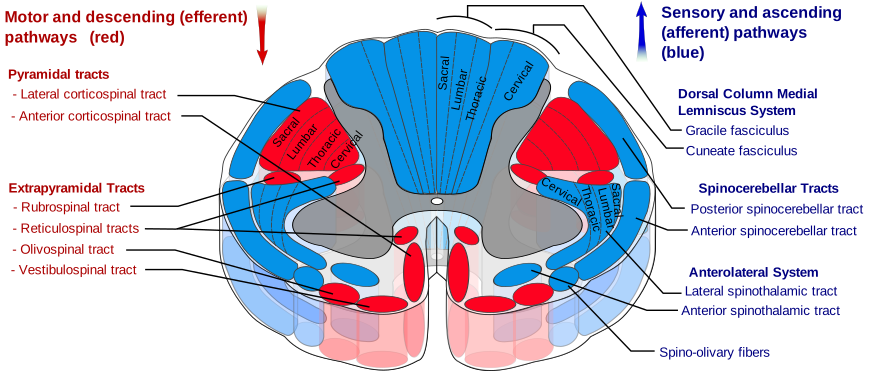
Somatotopy and blood supply (both are tested)
Lamination (somatotopy):
- Dorsal columns: legs medial (gracilis), arms lateral (cuneatus).
- Lateral corticospinal & spinothalamic tracts: medial → lateral = cervical, thoracic, lumbar, sacral. This is why central cord lesions hit the medial (arm/cervical) motor fibers and the midline crossing spinothalamic fibers first, and why the most lateral sacral fibers can be spared ("sacral sparing").
Arterial supply:
- One anterior spinal artery → anterior two-thirds (corticospinal, spinothalamic, anterior horns).
- Two posterior spinal arteries → posterior one-third (dorsal columns).
- Artery of Adamkiewicz (great anterior radicular artery), usually arising on the left, most often T9–T12 (range ~T8–L1), dominates the lower cord. The midthoracic cord is a watershed zone vulnerable to hypoperfusion (e.g., aortic cross-clamp/aneurysm repair).
Vignette: A man is stabbed in the back. Weeks later he has right-leg weakness with loss of position/vibration sense, and loss of pain/temperature in the left leg.
Below the lesion:
- Ipsilateral UMN weakness — lateral corticospinal (already crossed at the pyramids): spastic, hyperreflexic, Babinski.
- Ipsilateral loss of vibration/proprioception/fine touch — dorsal column (not yet crossed).
- Contralateral loss of pain/temperature — spinothalamic (crosses at the level), starting ~1–2 levels below.
At the level of the lesion:
- Ipsilateral LMN signs (flaccid, areflexic) from anterior horn/root damage.
- Ipsilateral loss of all sensation in a dermatomal band.
- If the lesion is above T1 → ipsilateral Horner syndrome (ptosis, miosis, anhidrosis) from interrupted descending sympathetics.

- Syringomyelia: central cavitation destroys crossing spinothalamic fibers at the anterior white commissure → bilateral, cape-like loss of pain/temperature (classically C8–T1) with dorsal columns spared. Linked to Chiari I; expansion reaches anterior horns → hand-muscle LMN atrophy. Patient painlessly burns the hands.
- Anterior spinal artery infarct: anterior two-thirds lost → bilateral motor + pain/temp loss below the lesion, vibration/proprioception preserved (dorsal columns spared). Classic after aortic repair; may begin as flaccid spinal shock then become UMN.
- Subacute combined degeneration (vitamin B12; also copper deficiency, N2O abuse): demyelination of dorsal columns + lateral corticospinal + spinocerebellar tracts → sensory ataxia, +Romberg, spastic paresis, paresthesias.
- Friedreich ataxia (AR; GAA trinucleotide repeat in frataxin/FXN): degeneration of dorsal columns + spinocerebellar + lateral corticospinal tracts (plus dorsal root ganglia) — same tracts as SCD but hereditary → mixed sensory + cerebellar ataxia, +Romberg, areflexia with an +Babinski, dysarthria, pes cavus/kyphoscoliosis; hypertrophic cardiomyopathy is the usual cause of death (also diabetes).
- Tabes dorsalis (tertiary neurosyphilis): degeneration of dorsal columns and dorsal roots → sensory ataxia, +Romberg, lancinating pains, absent DTRs, Argyll Robertson pupils, Charcot joints.
- ALS: combined UMN (lateral corticospinal) + LMN (anterior horn) degeneration, no sensory loss. Poliomyelitis and Werdnig-Hoffmann (SMA1) are pure LMN (anterior horn) → flaccid paralysis, no sensory loss.
Classic cord lesions at a glance
| Lesion | Structures affected | Key deficit | Classic association |
|---|---|---|---|
| Brown-Séquard (hemisection) | Ipsi dorsal column + corticospinal; contra spinothalamic | Ipsi motor + proprioception loss, contra pain/temp loss | Trauma/stab; Horner if above T1 |
| Syringomyelia | Crossing spinothalamic fibers (ant. white commissure) | Bilateral cape-like pain/temp loss; touch spared | Chiari I |
| Anterior spinal artery infarct | Anterior 2/3: corticospinal, spinothalamic, anterior horn | Bilateral motor + pain/temp loss; vibration spared | Aortic surgery, watershed |
| Subacute combined degeneration | Dorsal columns + lateral corticospinal + spinocerebellar | Sensory ataxia + spastic paresis + paresthesias | Vitamin B12 (also N2O, copper) |
| Friedreich ataxia | Dorsal columns + spinocerebellar + lateral corticospinal (+ DRG) | Ataxia, areflexia with +Babinski, pes cavus | AR GAA repeat (frataxin); HCM = cause of death |
| Tabes dorsalis | Dorsal columns + dorsal roots | Sensory ataxia, +Romberg, lancinating pain | Tertiary syphilis; Argyll Robertson pupils |
| ALS | Lateral corticospinal (UMN) + anterior horn (LMN) | Mixed UMN + LMN, no sensory loss | SOD1 (familial); riluzole |
| Polio / SMA (Werdnig-Hoffmann) | Anterior horn (LMN) | Flaccid paralysis, no sensory loss | Poliovirus; SMN1 (SMA) |
- UMN lesion: spastic paralysis, hyperreflexia, increased tone, Babinski present (upgoing toe), clonus, no early atrophy, no fasciculations.
- LMN lesion: flaccid paralysis, hyporeflexia/areflexia, decreased tone, downgoing/absent plantar, marked atrophy, fasciculations and fibrillations.
- Localize: weakness from a lesion above the pyramidal decussation = contralateral; in the cord = ipsilateral.
- ALS is the classic disease showing BOTH UMN and LMN signs with no sensory involvement.
- Crossing rule: *Dorsal columns and corticospinal cross in the medulla; spinothalamic crosses in the cord.* This one principle predicts every cord-lesion pattern.
- Argyll Robertson = "prostitute's pupil" — Accommodates but does not react (to light); flags tabes dorsalis / neurosyphilis.
- Dorsal columns carry "Fine touch, Vibration, Proprioception, Pressure" — the DCML modalities.
- Gracilis = Ground (legs), medial; Cuneatus = arms, lateral — dorsal-column somatotopy ("graceful legs").
- Subacute COMBINED degeneration = COMBINED tracts — dorsal columns + lateral corticospinal (+ spinocerebellar); think B12 (Friedreich hits the same tracts but is genetic).
Practice Anatomy now
Board-style questions, spaced-repetition flashcards, and a Socratic AI tutor — free to start.