Retinal Detachment & Vascular Occlusions
A high-yield Step 2 CK approach to sudden painless monocular vision loss, contrasting retinal detachment, CRAO, and CRVO by buzzwords, fundoscopy, and the next-best-step — including GCA workup and stroke-pathway management for CRAO.
The sudden painless vision loss cluster
Sudden, painless, monocular vision loss is the board's headline for this topic. Four heavy hitters cause it: retinal detachment (RD), central retinal artery occlusion (CRAO), central retinal vein occlusion (CRVO), and vitreous hemorrhage; amaurosis fugax is the transient warning shot.
Localize by the story:
- Flashes + floaters + a curtain/shadow advancing across the field → detachment.
- Instantaneous, profound loss with a pale retina + cherry-red spot → CRAO.
- Painless, variable loss with diffuse hemorrhages → CRVO.
Pain points away from these — think acute angle-closure glaucoma or optic neuritis instead. Your first two reflexes on the exam: check for a relative afferent pupillary defect (RAPD) and get dilated fundoscopy.
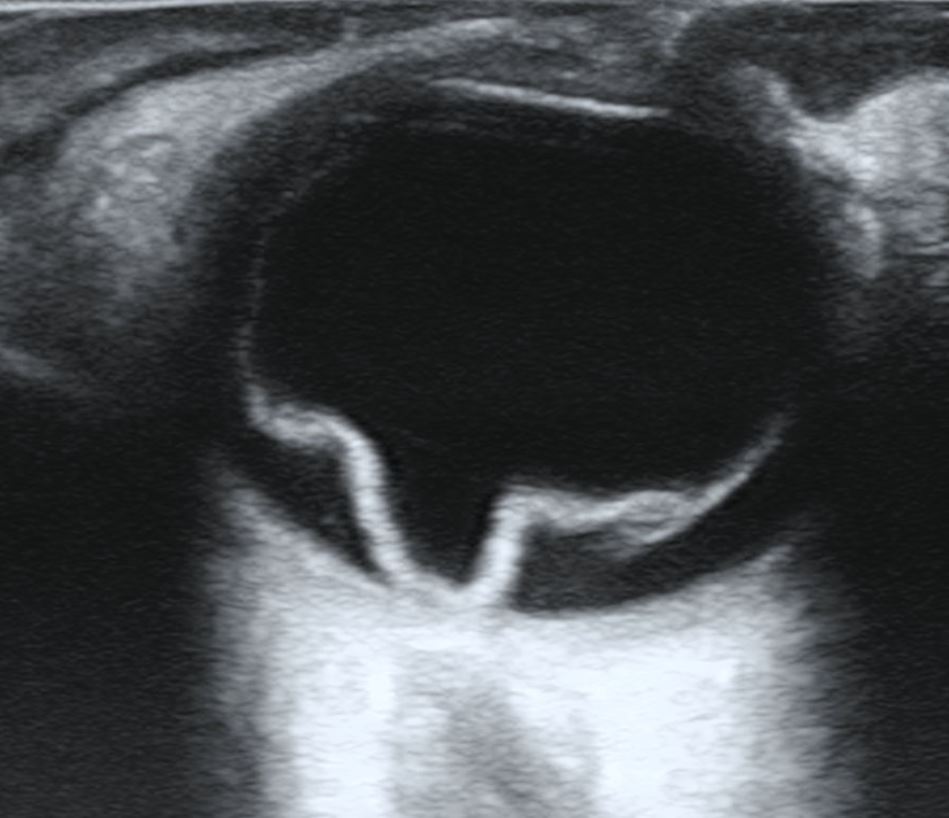
- Rhegmatogenous (most common): a retinal tear lets liquefied vitreous seep under the retina; usually follows a posterior vitreous detachment.
- Risk factors: high myopia, prior cataract/eye surgery (pseudophakia), trauma, lattice degeneration, older age.
- Classic sequence: photopsia (flashes) → new floaters ("cobwebs/soot") → a painless dark curtain/shadow advancing across the visual field.
- Fundoscopy: elevated, gray, wrinkled retina with a visible tear; red reflex may be diminished.
- Tractional RD: proliferative diabetic retinopathy, sickle cell — fibrous bands pull the retina off.
- Exudative/serous RD: no break — fluid from choroidal melanoma, malignant HTN, or inflammation.
- Mac-on (fovea still attached) = surgical emergency, repair within ~24 h to save central vision; mac-off = repair within days (fovea already off).
- Repair: laser/cryopexy seals tears; pneumatic retinopexy, scleral buckle, or vitrectomy reattach the retina.
- CRAO = "eye stroke": sudden, painless, profound monocular loss (often counting-fingers or worse) with a marked RAPD.
- Fundus: diffuse pale/white retina with a foveal cherry-red spot (fovea perfused by intact choroid); "boxcar" (cattle-truck) segmentation of the vessels.
- Irreversible retinal damage begins after roughly 90–100 min of complete occlusion → true emergency; manage on the acute stroke pathway (emergent referral, neuroimaging, embolic source workup).
- Sources: carotid atherosclerosis/embolus, cardiac embolus (AFib, valve). In age >50, suspect giant cell arteritis — check ESR/CRP, ask about headache, jaw claudication, scalp tenderness, and start high-dose corticosteroids empirically (don't wait for biopsy).
- BRAO: sectoral field loss; look for a Hollenhorst plaque (bright cholesterol embolus at a bifurcation).
- Amaurosis fugax: painless transient loss, a "curtain descending" for minutes that resolves = retinal TIA → carotid duplex, antiplatelet, vascular risk workup.
- Acute measures (low evidence): ocular massage, IOP-lowering (acetazolamide/timolol), anterior-chamber paracentesis.
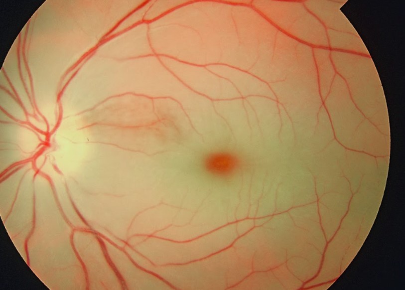
- CRVO: sudden, painless, variable monocular loss — often milder than CRAO, but severe if ischemic.
- Fundus: "blood and thunder" — diffuse retinal hemorrhages in all four quadrants, dilated tortuous veins, cotton-wool spots, and optic disc edema.
- Risk factors: HTN, diabetes, open-angle glaucoma, older age, hyperviscosity/hypercoagulable states.
- Subtypes: ischemic vs non-ischemic; the ischemic type carries high neovascularization risk.
- Complications: macular edema (main cause of vision loss) and neovascularization → neovascular glaucoma (iris/angle rubeosis).
- Management: anti-VEGF injections for macular edema; panretinal photocoagulation for neovascularization; control BP and glucose.
- BRVO: sectoral hemorrhages along one vein at an AV crossing, respecting the horizontal raphe; strongly tied to HTN; anti-VEGF for macular edema.
1. A 62-year-old myope reports a week of flashing lights and floaters, now a painless "gray curtain" crossing the lower field of the right eye. → Dx: rhegmatogenous retinal detachment. Next step: emergent same-day ophthalmology referral for dilated exam and surgical repair — establish mac-on vs mac-off.
2. A 74-year-old with atrial fibrillation has sudden, painless, total loss in one eye; fundus shows a pale retina with a cherry-red spot and a marked RAPD. → Dx: CRAO. Next step: treat as acute stroke (emergent evaluation, neuroimaging); check ESR/CRP and give high-dose steroids if any GCA features.
3. A 68-year-old hypertensive notes sudden painless blurring; fundus shows hemorrhages in all four quadrants with dilated tortuous veins. → Dx: CRVO. Next step: anti-VEGF for macular edema; screen for neovascularization; optimize BP and glucose.
The boards test these on pattern recognition — memorize the pairings:
- Cherry-red spot at the fovea (pale retina around it) → CRAO.
- "Blood and thunder" fundus → CRVO.
- Flashes + floaters + a curtain/shadow → retinal detachment.
- "Curtain descending" that lifts within minutes → amaurosis fugax (retinal TIA).
- Hollenhorst plaque (bright cholesterol embolus) → embolic/carotid arterial occlusion.
- Boxcar / cattle-truck vessel segmentation → arterial occlusion (CRAO).
- Painful red eye with a mid-dilated fixed pupil → not these — acute angle-closure glaucoma.
CRAO vs CRVO vs retinal detachment
| Feature | CRAO | CRVO | Rhegmatogenous RD |
|---|---|---|---|
| Onset | Instant | Hours | Hours–days |
| Vision loss | Profound | Variable | Curtain, then central |
| Prodrome | Amaurosis fugax | — | Flashes + floaters |
| Fundus | Pale retina, cherry-red spot, boxcar vessels | "Blood & thunder," tortuous dilated veins | Elevated gray retina + tear |
| RAPD | Marked | Mild; marked if ischemic | Mild/none |
| Workup | Stroke pathway; ESR/CRP for GCA | BP, glucose, hypercoag | Dilated exam; mac-on vs off |
| Treatment | Emergent stroke path; steroids if GCA | Anti-VEGF; PRP if neovascular | Retinopexy, scleral buckle, vitrectomy |
Practice Ophthalmology now
Board-style questions, spaced-repetition flashcards, and a Socratic AI tutor — free to start.