Respiratory Mechanics & V/Q Matching
A Step 1–focused walkthrough of respiratory mechanics (recoil balance, intrapleural/transpulmonary pressures, compliance, surfactant/Laplace) and V/Q matching (apex-vs-base gradient, HPV, shunt vs dead space, the alveolar gas equation and A–a gradient), anchored by NRDS and PE vignettes and a hypoxemia work-up table. Reviewed and verified against board-standard values (West-table V/Q numbers, alveolar gas math, HPV direction, CADET shifts) with the 100% O2 discriminating test as the load-bearing concept.
The Balance of Forces at Rest
Breathing is a tug-of-war between two elastic structures. The lung recoils inward (it wants to collapse); the chest wall recoils outward (it wants to spring open). At functional residual capacity (FRC) these opposing recoils are equal and opposite, so the respiratory system sits at equilibrium — airflow is zero and alveolar pressure = atmospheric (0 cmH2O).
The lung is held expanded against its recoil by a negative intrapleural pressure (~ −5 cmH2O at FRC). The net distending pressure holding an alveolus open is the transpulmonary pressure = alveolar − intrapleural = 0 − (−5) = +5 cmH2O.
- Inspiration: diaphragm contracts → thoracic volume ↑ → intrapleural pressure becomes more negative (~ −8) → alveolar pressure drops below atmospheric → air flows in.
- Expiration (quiet): passive — stored elastic recoil raises alveolar pressure above atmospheric → air flows out.
- Lose the negative intrapleural pressure (pneumothorax) → the lung collapses toward its unopposed recoil while the chest wall springs outward.
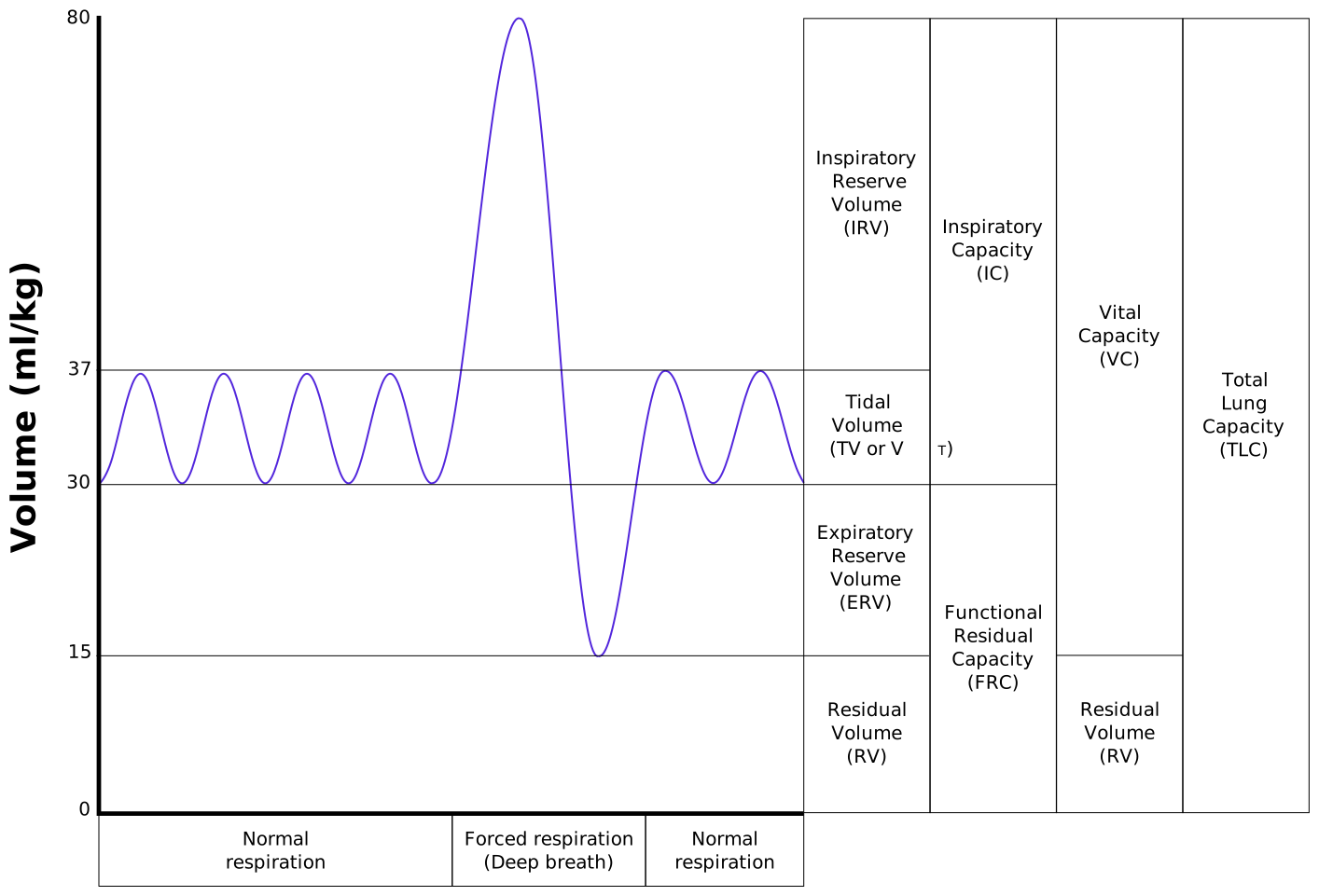
- Compliance = ΔV/ΔP — the ease of stretching the lung; it is the inverse of elastic recoil (compliance = 1/elastance). Lung compliance ≈ 200 mL/cmH2O (the whole lung–chest-wall system ≈ 100 mL/cmH2O).
- ↑ Compliance (floppy, low recoil): emphysema (elastin destruction) and normal aging → easy to fill, hard to empty → air trapping.
- ↓ Compliance (stiff, high recoil): pulmonary fibrosis, pulmonary edema, and surfactant deficiency (NRDS).
- Surfactant = dipalmitoylphosphatidylcholine (DPPC / lecithin), made by type II pneumocytes. It lowers alveolar surface tension → ↑ compliance and prevents atelectasis.
- Laplace's law: collapsing pressure P = 2T/r. Small alveoli (small r) generate a higher collapse pressure; surfactant lowers T disproportionately in small alveoli, stabilizing them so they don't empty into larger ones.
- Fetal lung maturity: surfactant synthesis begins ~week 20 and matures ~week 35; amniotic lecithin:sphingomyelin (L/S) ratio ≥ 2.0 indicates maturity.
Mechanism: deficient surfactant → ↑ surface tension → diffuse alveolar collapse (atelectasis) → ↓ compliance + collapsed-but-perfused alveoli (a physiologic shunt, V/Q → 0) → hypoxemia and ↑ work of breathing.
Vignette: a 28-week premature neonate — or an infant of a diabetic mother (fetal hyperinsulinemia antagonizes cortisol and delays surfactant) or a baby delivered by C-section without labor — develops tachypnea, grunting, nasal flaring, and intercostal retractions within hours of birth. CXR shows a diffuse ground-glass / reticulogranular pattern with air bronchograms.
Prevent / Treat: antenatal maternal glucocorticoids (betamethasone) accelerate fetal surfactant; postnatal exogenous surfactant + CPAP/O2. Watch for O2/ventilation complications — retinopathy of prematurity and bronchopulmonary dysplasia.
Why V/Q Differs Top to Bottom
Gravity makes both ventilation and perfusion greater at the base than the apex — but perfusion increases more steeply from apex to base than ventilation does. The result is a regional gradient in the ventilation/perfusion (V/Q) ratio:
- Apex → high V/Q (~3): relatively over-ventilated. Alveolar gas approaches inspired air — high PO2 (~132), low PCO2 (~28). The oxygen-rich, low-flow apex is why reactivation TB (an obligate aerobe) and apical bullae favor the upper lobes.
- Base → low V/Q (~0.6): relatively over-perfused. Gas approaches venous values — lower PO2 (~89), higher PCO2 (~42).
- Whole-lung average V/Q ≈ 0.8 (alveolar ventilation ~4 L/min ÷ cardiac output ~5 L/min).
Hypoxic pulmonary vasoconstriction (HPV): unlike every systemic bed, pulmonary arterioles constrict when alveolar PO2 falls, shunting blood away from poorly ventilated regions toward well-ventilated ones to preserve V/Q matching. Chronic global hypoxia (COPD, high altitude) → diffuse HPV → pulmonary hypertension → cor pulmonale.
Lung Apex vs Base (Upright, at Rest)
| Feature | Apex | Base |
|---|---|---|
| Ventilation | Lower | Higher |
| Perfusion (blood flow) | Lowest | Highest |
| V/Q ratio | High (~3) | Low (~0.6) |
| Alveolar PO2 | High (~132) | Lower (~89) |
| Alveolar PCO2 | Low (~28) | Higher (~42) |
| pH of end-capillary blood | Higher | Lower |
| Physiologic trend | Toward "wasted ventilation" (dead space) | Toward "wasted perfusion" (shunt) |
| Disease association | Reactivation TB, apical bullae, centriacinar (smoking) emphysema | Panacinar (α1-antitrypsin) emphysema, basal edema |
- V/Q = 0 → SHUNT (perfusion, no ventilation): airway obstruction, atelectasis, lobar collapse. Blood retains mixed-venous values (PO2 40, PCO2 46). Hypoxemia does NOT correct with 100% O2.
- V/Q = ∞ → DEAD SPACE (ventilation, no perfusion): pulmonary embolism. Alveolar gas approaches inspired air (PO2 150, PCO2 0).
- Alveolar gas equation: PAO2 = FiO2·(Patm − PH2O) − PaCO2/R = 0.21·(760 − 47) − 40/0.8 ≈ 150 − 50 = 100 mmHg (room air, sea level).
- A–a gradient = PAO2 − PaO2; normal ~5–15 mmHg, rises with age (≈ age/4 + 4).
- ↑ A–a gradient: V/Q mismatch, right-to-left shunt, diffusion limitation.
- Normal A–a gradient: high altitude and hypoventilation (opioids, obesity-hypoventilation) — the alveolar–arterial transfer is intact; the driving PAO2 is simply low.
- 100% O2 test: improves V/Q mismatch & diffusion limitation, but fails to correct a true shunt — the discriminating step.
Mechanism: a clot occludes a pulmonary artery branch → those alveoli are ventilated but not perfused → physiologic dead space (V/Q → ∞). Dead space alone doesn't drop PaO2 — the hypoxemia comes from reflex (hypocapnic) bronchoconstriction, surfactant loss/atelectasis, and blood redistributed into overperfused units, i.e. V/Q mismatch and shunt with an ↑ A–a gradient; the patient hyperventilates → respiratory alkalosis (low PaCO2).
Vignette: a post-op patient, long-haul traveler, or OCP user with sudden pleuritic chest pain, dyspnea, and tachycardia; SpO2 low, PaCO2 low. ECG usually shows sinus tachycardia (the classic but uncommon finding is S1Q3T3). CT pulmonary angiography is the gold standard.
Contrast — right-to-left shunt (Eisenmenger, ARDS, complete atelectasis): also ↑ A–a gradient, but hypoxemia is refractory to 100% O2 — the bedside way to separate shunt from dead space / mismatch.
"CADET, face Right!" — factors that shift the O2–hemoglobin curve to the RIGHT (↓ affinity, ↑ P50, more O2 unloaded to tissues):
- C — ↑ CO2
- A — Acid / ↓ pH (Bohr effect)
- D — ↑ 2,3-DPG (2,3-BPG)
- E — Exercise
- T — ↑ Temperature
Left shift (↑ affinity, holds O2): the opposites — ↓ 2,3-BPG, alkalosis, ↓ temperature — plus fetal Hb (HbF), carbon monoxide (CO), and methemoglobin.
Shape: hemoglobin's curve is sigmoidal (cooperative binding across 4 subunits); myoglobin is hyperbolic and left-shifted (1 subunit, high affinity → tissue O2 storage).
V/Q anchors: "Zero is a shunt — 100% O2 won't fix it. Infinity is dead space — think embolus."

Causes of Hypoxemia — A–a Gradient & O2 Response
| Cause | A–a gradient | Corrects with 100% O2? | Classic example |
|---|---|---|---|
| High altitude | Normal | Yes | Mountaineer (↓ inspired PO2) |
| Hypoventilation | Normal | Yes | Opioid overdose, obesity-hypoventilation |
| V/Q mismatch | ↑ | Yes (largely) | COPD, asthma, PE |
| Diffusion limitation | ↑ | Yes | Fibrosis, emphysema, exertion |
| Right-to-left shunt | ↑ | No | Eisenmenger, ARDS, atelectasis |
Practice Physiology now
Board-style questions, spaced-repetition flashcards, and a Socratic AI tutor — free to start.