Respiratory Histology
A boards-focused walk through respiratory histology: the conducting-to-respiratory epithelial gradient, key cells (goblet, club, type I/II pneumocytes, macrophages, neuroendocrine), and the classic vignettes of neonatal RDS and Kartagener syndrome.
How the boards frame respiratory histology
The respiratory tract splits into a conducting zone (nose → terminal bronchioles) that warms, humidifies, and filters air but does no gas exchange, and a respiratory zone (respiratory bronchioles → alveoli) where gas exchange happens. The single most tested idea is the epithelial gradient: pseudostratified ciliated columnar ("respiratory epithelium") with goblet cells in trachea/bronchi → simple ciliated columnar/cuboidal in bronchioles → simple squamous (type I pneumocytes) in alveoli. Moving distally, four things disappear roughly in order — cartilage, submucosal glands, goblet cells, then cilia — while smooth muscle becomes relatively prominent (why bronchioles constrict in asthma). The mucociliary escalator (cilia sweeping trapped mucus up toward the pharynx) is the key conducting-airway defense; alveoli have no cilia or goblet cells and rely on macrophages to clear debris.
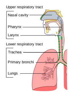
- Respiratory epithelium = pseudostratified ciliated columnar + goblet cells; cilia sweep mucus toward the pharynx
- Goblet cells are gone by the terminal bronchiole; cilia extend one step further, to the respiratory bronchioles — cilia must sit distal to goblet cells so mucus is never stranded
- Bronchioles: NO cartilage, NO glands, NO goblet cells but prominent smooth muscle → site of constriction in asthma
- Club (Clara) cells: nonciliated, dome-shaped; secrete a component of surfactant, detoxify via CYP450, and act as bronchiolar stem cells
- Type I pneumocytes: simple squamous, ~97% of alveolar surface, gas exchange, cannot divide
- Type II pneumocytes: cuboidal, lamellar bodies, make surfactant, and are the stem cell regenerating both type I and II after injury
- Alveolar macrophages ("dust cells"): hemosiderin-laden = "heart-failure cells" in CHF/pulmonary edema
- Neuroendocrine (Kulchitsky) cells → origin of small cell carcinoma and carcinoid
Airway transitions (what changes as you descend)
| Level | Epithelium | Cartilage | Goblet cells | Notable |
|---|---|---|---|---|
| Trachea / bronchi | Pseudostratified ciliated columnar | Yes (rings → plates) | Yes | Submucosal glands |
| Bronchioles | Simple ciliated columnar → cuboidal | None | Few → none | Smooth muscle prominent |
| Terminal bronchiole | Simple cuboidal, ciliated | None | None | Club cells; last conducting segment |
| Respiratory bronchiole | Simple cuboidal + scattered alveoli | None | None | First gas exchange |
| Alveolus | Simple squamous (type I) | None | None | Type II cells + macrophages |

Vignette: A 28-week premature neonate develops tachypnea, grunting, nasal flaring, and intercostal retractions within hours of birth. CXR shows diffuse ground-glass (reticulogranular) opacities with air bronchograms. Amniotic lecithin:sphingomyelin (L:S) ratio was < 2.
Diagnosis: Neonatal respiratory distress syndrome (RDS) — deficient surfactant from immature type II pneumocytes. Surfactant (chiefly dipalmitoylphosphatidylcholine, DPPC) lowers alveolar surface tension and prevents end-expiratory collapse; deficiency → diffuse atelectasis.
Risk factors: prematurity, maternal diabetes (fetal hyperinsulinemia delays maturation), C-section without labor.
Next best step: respiratory support (CPAP/ventilation) + exogenous surfactant; give antenatal betamethasone to the mother when preterm delivery is anticipated (prevention — accelerates surfactant synthesis). Watch for oxygen/ventilation complications: retinopathy of prematurity and bronchopulmonary dysplasia.
Type I vs Type II pneumocytes
| Feature | Type I pneumocyte | Type II pneumocyte |
|---|---|---|
| Shape | Simple squamous (thin) | Cuboidal |
| Numbers vs area | ~40% of cells, ~97% of surface | ~60% of cells, small surface area |
| Function | Gas exchange (blood–air barrier) | Surfactant (lamellar bodies) |
| Regeneration | Cannot divide | Stem cell → replaces I & II |
| Clinical link | Injured in diffuse alveolar damage / ARDS | Deficient in neonatal RDS; proliferate after injury |
- Lamellar bodies → surfactant stored in type II pneumocytes
- L:S (lecithin:sphingomyelin) ratio ≥ 2:1 → mature lungs; < 2 → risk of neonatal RDS (lecithin = phosphatidylcholine; DPPC is the main surfactant lecithin)
- "Heart-failure cells" → hemosiderin-laden alveolar macrophages in CHF/edema
- "Dust cells" → alveolar macrophages
- Kulchitsky / neuroendocrine cells → small cell carcinoma & carcinoid
- Type II = 2 jobs → makes surfactant and is the alveolar stem cell
Vignette: A young adult has chronic sinusitis, recurrent otitis media, bronchiectasis, and reduced fertility (immotile sperm in men; dysfunctional fallopian-tube cilia with ectopic-pregnancy risk in women). A routine CXR shows dextrocardia, and imaging confirms situs inversus.
Diagnosis: Primary ciliary dyskinesia (Kartagener syndrome) — a defect in the axonemal dynein arms of cilia. Without functional cilia on the respiratory epithelium, the mucociliary escalator fails → recurrent sinopulmonary infections and bronchiectasis; dysfunctional embryonic nodal cilia → ~50% have situs inversus.
Buzzword triad: situs inversus + chronic sinusitis + bronchiectasis. Next step: screen with low nasal nitric oxide; confirm with ciliary electron microscopy / genetics. This is exactly why cilia on respiratory epithelium are high-yield.
Practice Histology now
Board-style questions, spaced-repetition flashcards, and a Socratic AI tutor — free to start.