Respiratory & Lung Development
A Step 1 embryology lesson tracing the respiratory tree from the week-4 laryngotracheal diverticulum through the five stages of lung development, with the germ-layer derivatives, surfactant biology, and the board-favorite congenital defects (TEF/esophageal atresia, congenital diaphragmatic hernia, neonatal RDS, Potter-related pulmonary hypoplasia). Emphasis is on what goes wrong developmentally and how each malformation presents.
Origin & Branching of the Airway
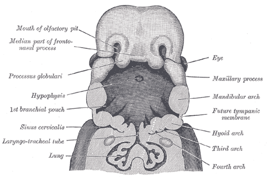
The respiratory system is a foregut derivative. Around week 4, the respiratory (laryngotracheal) diverticulum buds off the ventral wall of the foregut, just caudal to the pharyngeal pouches. Its endodermal lining gives rise to the epithelium and glands of the larynx, trachea, bronchi, and the pulmonary alveoli; the surrounding splanchnic (visceral) mesoderm forms the cartilage, smooth muscle, connective tissue, pulmonary vasculature, and visceral pleura.
The diverticulum is separated from the dorsal esophagus by the tracheoesophageal (TE) septum, formed as paired tracheoesophageal folds fuse in the midline — partitioning the foregut into a ventral respiratory tube and a dorsal esophagus. Faulty partitioning here is the basis of tracheoesophageal fistula.
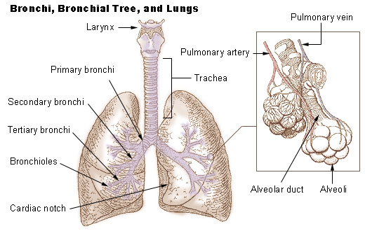
The growing lung bud divides into two bronchial buds → primary (main) bronchi, then secondary (lobar) bronchi — 3 on the right, 2 on the left (matching the lung lobes) — and tertiary (segmental) bronchi that define the bronchopulmonary segments (~10 per lung). The more vertical right main bronchus is why aspirated material preferentially enters the right lung. Repeated branching morphogenesis, driven by epithelial–mesenchymal signaling, then builds the full conducting and respiratory tree.
- Epithelium/glands of larynx → alveoli = endoderm; cartilage/muscle/vessels/pleura = splanchnic mesoderm.
- Lung arises from the ventral foregut as the laryngotracheal diverticulum (~week 4).
- The TE septum separates trachea (ventral) from esophagus (dorsal); its failure → TEF / esophageal atresia.
- Lobar bronchi: Right = 3, Left = 2; the right main bronchus is more vertical → aspiration favors the right lung.
- Surfactant is made by type II pneumocytes; main component is dipalmitoylphosphatidylcholine (lecithin).
- L:S (lecithin:sphingomyelin) ratio ≥ 2 = lung maturity; surfactant deficiency → neonatal RDS.
- Gas-exchange viability begins in the canalicular stage (~week 25), once pneumocytes differentiate and capillaries invest the air spaces.
Five Stages of Lung Development
| Stage | Weeks | Key developmental events | Clinical correlate |
|---|---|---|---|
| Embryonic | 4–7 | Lung bud → trachea; bronchial buds → main / lobar / segmental bronchi | TEF, pulmonary agenesis |
| Pseudoglandular | 5–17 | Branching down to terminal bronchioles; no gas-exchange structures yet | Birth here = nonviable (respiration impossible); CPAM |
| Canalicular | 16–26 | Terminal bronchioles → respiratory bronchioles → alveolar ducts; capillaries proliferate; type I & II pneumocytes differentiate | Respiration possible ~week 25 |
| Saccular | 26–birth | Terminal sacs form (separated by primary septa); surfactant secreted; air–blood barrier thins | Marginal viability with support |
| Alveolar | 36 wk–~8 yr | Secondary septation → true alveoli multiply | Most alveoli form after birth (~20–50 million at birth → ~300 million+ in adult) |

Surfactant & the Fetal-to-Neonatal Transition
Surfactant — a phospholipid–protein film (chiefly dipalmitoylphosphatidylcholine / lecithin, plus phosphatidylglycerol and surfactant proteins A–D) secreted by type II pneumocytes — lowers alveolar surface tension, preventing end-expiratory collapse (atelectasis) and raising compliance (by Laplace, small alveoli would otherwise empty into larger ones). Synthesis begins around week 24 as type II pneumocytes mature, but reaches reliably adequate levels only around week 35; an amniotic L:S ratio ≥ 2 and the presence of phosphatidylglycerol confirm maturity.
In utero the lungs are fluid-filled and pulmonary vascular resistance is high (blood shunts right→left through the ductus arteriosus and foramen ovale). At birth, aeration plus surfactant drop alveolar surface tension, pulmonary vascular resistance falls, and rising O₂ with increased left atrial pressure closes the fetal shunts.
Surfactant deficiency → Neonatal Respiratory Distress Syndrome (hyaline membrane disease): a preterm infant with tachypnea, grunting, nasal flaring, retractions, and cyanosis, and a CXR showing diffuse ground-glass opacities with air bronchograms. Risks: prematurity, maternal diabetes (fetal hyperinsulinemia antagonizes cortisol-driven surfactant synthesis), and cesarean delivery without labor. Antenatal glucocorticoids (betamethasone) accelerate production; cortisol and thyroxine stimulate it, while insulin inhibits.
A newborn develops coughing, choking, and cyanosis with the first feeding and has excessive drooling. A nasogastric tube coils in the upper chest and cannot be advanced. The pregnancy was complicated by polyhydramnios, and an abdominal film shows a gastric air bubble.
- Diagnosis: esophageal atresia with distal tracheoesophageal fistula — the most common variant (~85%): the proximal esophagus ends blind while the distal esophagus connects to the trachea (air enters the stomach; feeds are aspirated).
- Pure esophageal atresia (no fistula) → gasless abdomen; isolated "H-type" TEF presents later with recurrent aspiration pneumonia.
- Screen for the VACTERL association.
- Mechanism: deviation/failure of the tracheoesophageal septum during the embryonic stage.
A term neonate is cyanotic and tachypneic at birth with a scaphoid (sunken) abdomen. Breath sounds are diminished on the left, and bowel sounds are audible in the left chest. CXR shows bowel loops in the left hemithorax with the mediastinum shifted to the right.
- Diagnosis: congenital diaphragmatic hernia from failure of the pleuroperitoneal membrane(s) to close, allowing abdominal viscera to herniate into the thorax and compress the developing lung → pulmonary hypoplasia (the main driver of morbidity and mortality — the problem is the hypoplastic lung, not the hernia itself).
- Most are posterolateral and left-sided = Bochdalek; the rarer anterior (parasternal/retrosternal) form is Morgagni.
Congenital Respiratory Defects — Mechanism → Presentation
| Defect | Developmental mechanism | Classic presentation |
|---|---|---|
| Esophageal atresia + distal TEF | Faulty TE septum partitioning of the foregut | Drooling, choking/cyanosis with feeds, NG tube coils, polyhydramnios, gastric air bubble; VACTERL |
| Congenital diaphragmatic hernia (Bochdalek) | Pleuroperitoneal membrane fails to close (usually left posterolateral) | Scaphoid abdomen, bowel in chest, mediastinal shift, pulmonary hypoplasia |
| Neonatal RDS | Surfactant deficiency (immature type II pneumocytes) | Preterm; grunting, retractions; CXR ground-glass + air bronchograms |
| Pulmonary hypoplasia | Oligohydramnios (Potter) or thoracic compression (e.g., CDH) | Small underdeveloped lungs, respiratory failure; part of Potter sequence |
| Bronchopulmonary sequestration | Accessory lung bud; nonfunctional tissue with no airway connection, fed by a systemic (aortic) artery | Recurrent infection or incidental mass; lacks normal bronchial communication |
| CPAM / CCAM | Abnormal pseudoglandular-stage airway patterning → hamartomatous cysts | Cystic lung lesion; respiratory distress or recurrent infection |
- VACTERL (TEF associations): Vertebral defects, Anal atresia, Cardiac defects, Tracheo-Esophageal fistula, Renal anomalies, Limb (radial) defects.
- CDH sides — "Bochdalek is Back and to the Left": Bochdalek = Back (posterolateral), usually left, and more common; Morgagni = anterior/parasternal (Medial), rarer.
- Potter sequence — "POTTER": Pulmonary hypoplasia, Oligohydramnios (the cause), Twisted face (Potter facies), Twisted skin, Extremity/limb defects, Renal agenesis (bilateral). Oligohydramnios → lungs cannot expand → pulmonary hypoplasia.
- NRDS risk ↑: prematurity, maternal diabetes (fetal hyperinsulinemia ↓ surfactant), C-section without labor, male sex.
- NRDS complications: PDA; from O₂/ventilation — retinopathy of prematurity and bronchopulmonary dysplasia; also NEC and IVH.
- Antenatal betamethasone ↑ fetal surfactant; cortisol/thyroxine stimulate, insulin inhibits.
- CDH → pulmonary hypoplasia is the lethal problem — not the hernia itself.
- Oligohydramnios (Potter) → pulmonary hypoplasia; conversely, polyhydramnios suggests esophageal atresia (the fetus cannot swallow amniotic fluid).
- Born in the pseudoglandular stage = nonviable (no gas-exchange surface); surfactant + capillary investment by the saccular stage allow survival with support.
Practice Embryology now
Board-style questions, spaced-repetition flashcards, and a Socratic AI tutor — free to start.