Renal Tubular Acidosis
A Step 1-focused lesson on renal tubular acidosis that frames all types as a normal anion gap (hyperchloremic) metabolic acidosis and teaches the board discriminators — serum potassium, urine pH, and the urine anion gap — through a comparison table, classic vignettes, and next-best-step management. Corrected the urine anion gap teaching: it is positive in distal (Type 1) and Type 4 RTA but unreliable (often negative) in proximal (Type 2) RTA, which is identified by its Fanconi features instead.
The big picture
Renal tubular acidosis (RTA) is a group of tubular disorders producing a normal anion gap (hyperchloremic) metabolic acidosis with a normal or near-normal GFR — the defect is in tubular handling of acid/bicarbonate, not in glomerular filtration. Physiologically, the proximal tubule reclaims ~80% of filtered HCO3⁻, and the α-intercalated cells of the collecting duct secrete H⁺, regenerating HCO3⁻ and excreting acid buffered as titratable acid and NH4⁺.
Boards test three types:
- Type 2 (proximal) — failure to reabsorb HCO3⁻.
- Type 1 (distal) — failure to secrete H⁺.
- Type 4 (hyperkalemic) — aldosterone deficiency or resistance.
(Type 3 is a rare combined form and is essentially never tested.) The fastest way to separate them on a vignette is the serum potassium and the urine pH.
- Every RTA = normal anion gap metabolic acidosis (↑Cl⁻). A normal gap is what separates RTA from lactic acidosis, DKA, uremia, and toxins.
- Type 1 (distal): can't secrete H⁺ → urine pH > 5.5 (inappropriately alkaline), hypokalemia, calcium-phosphate stones + nephrocalcinosis. Triggers: Sjögren / SLE / RA, amphotericin B, congenital.
- Type 2 (proximal): wastes HCO3⁻ → urine pH usually < 5.5 at steady state (distal acidification is intact), hypokalemia, part of Fanconi syndrome (glucosuria, phosphaturia, aminoaciduria, uricosuria). Triggers: multiple myeloma, tenofovir, acetazolamide, Wilson disease, cystinosis, lead.
- Type 4 (hyperkalemic): ↓aldosterone effect → hyperkalemia suppresses ammoniagenesis → low NH4⁺ buffer, urine pH < 5.5. Triggers: diabetic nephropathy (hyporeninemic hypoaldosteronism), ACEi/ARB, NSAIDs, heparin, spironolactone, trimethoprim.
- Only Type 4 is hyperkalemic; Types 1 and 2 are hypokalemic.

Side-by-side comparison
| Feature | Type 1 (Distal) | Type 2 (Proximal) | Type 4 (Hyperkalemic) |
|---|---|---|---|
| Defect | ↓ H⁺ secretion (α-intercalated cell) | ↓ HCO3⁻ reabsorption (proximal tubule) | ↓ aldosterone (deficiency/resistance) |
| Serum K⁺ | Low | Low | High |
| Urine pH | > 5.5 (can't acidify) | Usually < 5.5 (steady state) | < 5.5 |
| Serum HCO3⁻ | Severe (may be < 10) | Moderate (~12–18) | Mild (> 15) |
| Urine anion gap | Positive (↓NH4⁺) | Unreliable — often negative (distal NH4⁺ intact) | Positive (↓NH4⁺) |
| Stones / bone | Ca-phosphate stones, nephrocalcinosis | Osteomalacia / rickets; no stones | — |
| Classic causes | Sjögren, SLE, amphotericin B | Fanconi: myeloma, tenofovir, acetazolamide, Wilson | Diabetic nephropathy, ACEi/ARB, NSAIDs, trimethoprim |
| Treatment | Potassium citrate (low-dose alkali) | High-dose HCO3⁻ + K⁺, thiazide | Fludrocortisone, low-K⁺ diet, loop diuretic |
Approach any normal anion gap metabolic acidosis like this:
- Confirm the normal gap (Na − Cl − HCO3⁻ ≈ 8–12) with hyperchloremia.
- Separate renal from GI (diarrhea) loss with the urine anion gap: UAG = U_Na + U_K − U_Cl, a surrogate for urinary NH4⁺.
- Negative UAG → abundant NH4⁺ excreted = kidney responding appropriately = GI/diarrhea (proximal RTA can also read negative, because distal ammonium handling is intact).
- Positive UAG → low NH4⁺ = impaired distal acid excretion = distal (Type 1) or Type 4 RTA.
- Caveat: the UAG is validated to distinguish distal RTA/Type 4 from GI losses; it is unreliable in proximal (Type 2) RTA, which is instead recognized by its Fanconi features.
- Localize with serum K⁺ (high → Type 4; low → Type 1 or 2) and urine pH (persistently > 5.5 despite acidemia → Type 1).
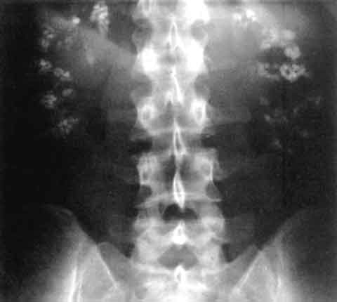
- RTA is a clinical/biochemical diagnosis — biopsy is not used to diagnose it. Imaging may show nephrocalcinosis (Type 1); look for glucosuria with a normal serum glucose (Fanconi → Type 2).
Vignette: A 38-year-old woman with dry eyes and dry mouth (Sjögren syndrome) has fatigue and muscle weakness. Labs: non-anion-gap metabolic acidosis, HCO3⁻ 12, K⁺ 2.9. Despite the acidemia, urine pH is 6.5. Renal ultrasound shows medullary nephrocalcinosis.
- Diagnosis: Type 1 (distal) RTA. The give-away is an inappropriately alkaline urine (pH > 5.5) in the face of systemic acidemia; hypokalemia + nephrocalcinosis + Sjögren seal it.
- Next best step / management: Oral alkali as potassium citrate — corrects the acidosis, replaces K⁺, and raises urinary citrate to prevent calcium-phosphate stones/nephrocalcinosis. Prefer potassium citrate over plain sodium bicarbonate, which can acutely worsen the hypokalemia.
Vignette: A 64-year-old man with type 2 diabetes and CKD taking lisinopril has K⁺ 5.9 and a mild non-anion-gap acidosis (HCO3⁻ 18). Urine pH is 5.0.
- Diagnosis: Type 4 RTA from hyporeninemic hypoaldosteronism (classic in diabetic nephropathy). The pairing of hyperkalemia with an acidic urine and only mild acidosis is the tell — the hyperkalemia itself suppresses ammoniagenesis, limiting NH4⁺ buffer even though the H⁺ pump works.
- Next best step: Stop/reduce the ACE inhibitor and other K⁺-raising drugs, start a low-potassium diet ± a loop diuretic; add fludrocortisone when there is true hypoaldosteronism without volume overload. If K⁺ is dangerously high or there are ECG changes, treat the hyperkalemia first.
- "neGUTive" urine anion gap = GI (gut) cause → diarrhea. A positive UAG points to impaired renal acid excretion → distal (Type 1) or Type 4 RTA (not reliable for proximal RTA).
- Potassium by number: Types 1 & 2 → low K⁺; Type 4 → high K⁺ ("4 = more potassium").
- Proximal = Type 2 = Fanconi, which wastes everything up front: glucose, phosphate, amino acids, uric acid, and HCO3⁻.
- Type 1 is the "one" that can't acidify — the only type with urine pH > 5.5 despite acidemia.
- Type 1 (distal): Potassium citrate (or sodium bicarbonate) — low-dose alkali usually suffices; citrate also counters the stone/nephrocalcinosis risk. Correcting acidosis improves growth and bone disease in children.
- Type 2 (proximal): Needs large doses of alkali (bicarbonate is continuously wasted) plus generous K⁺ (alkali worsens the hypokalemia); a thiazide induces mild volume contraction that enhances proximal reabsorption. Add phosphate/vitamin D for osteomalacia and treat the cause (stop tenofovir, treat myeloma).
- Type 4 (hyperkalemic): Lower the K⁺ — low-potassium diet, loop diuretic, and stop offending drugs (ACEi/ARB, NSAIDs, K⁺-sparing diuretics, trimethoprim); fludrocortisone replaces mineralocorticoid when there is genuine hypoaldosteronism.
Practice Renal now
Board-style questions, spaced-repetition flashcards, and a Socratic AI tutor — free to start.