Pulmonary Hypertension & Cor Pulmonale
A Step 1–2 walk through pulmonary hypertension and cor pulmonale — pathophysiology (PVR, remodeling, plexiform lesions), the 5 WHO groups, echo-then-right-heart-cath diagnosis, drug-suffix pathways, and group-specific management. Two board-classic vignettes (idiopathic PAH vs COPD cor pulmonale) drill the pre- vs postcapillary distinction and next-best-step decisions. Reviewed and verified against 2022 ESC/ERS thresholds; no mechanism, PFT/imaging, or next-best-step errors found.
Pathophysiology: from high resistance to a failing RV
Pulmonary hypertension (PH) = sustained rise in mean pulmonary arterial pressure (mPAP). The traditional cutoff is ≥25 mmHg at rest by right heart catheterization; the 2022 ESC/ERS guidelines lowered it to >20 mmHg. Precapillary PH additionally requires PAWP ≤15 mmHg and PVR >2 Wood units.
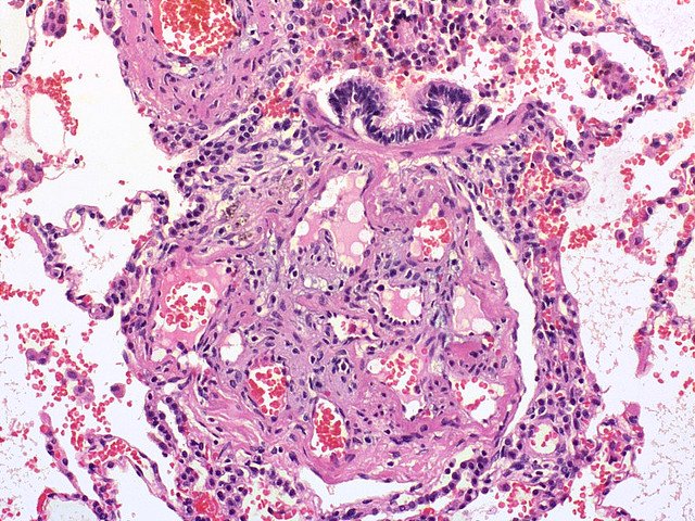
The engine is a rise in pulmonary vascular resistance (PVR) from three processes: vasoconstriction, vascular remodeling (medial smooth-muscle hypertrophy, intimal fibrosis, and — in advanced PAH — plexiform lesions), and in-situ thrombosis. Endothelial dysfunction tips the balance toward vasoconstrictors (endothelin-1, thromboxane) and away from vasodilators (NO, prostacyclin).
Chronically high PVR raises RV afterload → RV hypertrophy → dilation → failure. Cor pulmonale is RV enlargement/failure caused by a primary pulmonary (lung parenchymal or vascular) disorder — by definition it excludes RV failure that is secondary to left heart disease.
- Group 1 — PAH (precapillary): idiopathic; heritable (BMPR2 loss-of-function); drugs/toxins (anorexigens/fen-phen, methamphetamine); connective tissue disease (scleroderma/CREST = #1 CTD cause); HIV; portal hypertension (portopulmonary); congenital L→R shunts (may reverse to Eisenmenger); schistosomiasis (most common cause of PAH worldwide)
- Group 2 — Left heart disease (MOST COMMON cause of PH overall; postcapillary): HFrEF, HFpEF, mitral stenosis/regurgitation → PAWP >15
- Group 3 — Lung disease / hypoxia: COPD, ILD, OSA, chronic high-altitude → hypoxic pulmonary vasoconstriction
- Group 4 — CTEPH: chronic organized/unresolved PE; potentially curable surgically
- Group 5 — Multifactorial/unclear: sarcoidosis, myeloproliferative/hematologic disease, metabolic disorders, chronic renal failure
Board reflex: young woman + exertional dyspnea + loud P2 = suspect Group 1 PAH; older patient with orthopnea/HFpEF = Group 2 (the commonest cause overall).
Precapillary vs postcapillary (PAWP is the splitter)
| Feature | Precapillary (Groups 1, 3, 4) | Postcapillary (Group 2) |
|---|---|---|
| mPAP | >20 mmHg | >20 mmHg |
| PAWP | ≤15 mmHg | >15 mmHg |
| PVR | >2 Wood units | ≤2 (isolated postcap) |
| Problem site | pulmonary arteries / lung | left heart |
| Classic causes | IPAH, COPD/ILD, CTEPH | HFrEF/HFpEF, mitral disease |
| PAH-specific drugs | may help (Group 1 only) | contraindicated (pulmonary edema) |
A 32-year-old woman has 8 months of progressive exertional dyspnea and fatigue, plus one episode of syncope while climbing stairs. Exam: loud, palpable P2, a left parasternal (RV) heave, a holosystolic murmur at the left lower sternal border that increases with inspiration (tricuspid regurgitation, Carvallo sign), elevated JVP with a prominent a-wave (forceful RA contraction against a stiff, hypertrophied RV), and pedal edema. Lungs are clear. ECG: right-axis deviation and RVH.
- Diagnosis: idiopathic pulmonary arterial hypertension (Group 1) — clear lungs + right-heart signs point away from parenchymal disease.
- Next best step: transthoracic echocardiography (best initial/screening test) to estimate PASP and assess the RV.
- Then confirm: right heart catheterization (gold standard) with acute vasoreactivity testing.
Buzzword pearl: exertional syncope = poor prognosis (fixed, non-augmentable cardiac output).
- Echocardiography = best initial/screening test: estimates PASP from the TR jet velocity; shows RV dilation/hypertrophy and a D-shaped septum (LV flattening) from RV pressure overload
- Right heart catheterization (Swan-Ganz) = GOLD STANDARD / confirmatory: directly measures mPAP, PAWP, PVR, CO → separates pre- vs postcapillary
- Acute vasoreactivity testing (inhaled NO, IV adenosine, or IV/inhaled prostacyclin) during RHC: positive → trial of a CCB
- ECG: right-axis deviation, RVH, P pulmonale (tall peaked P in II), RV strain (TWI V1–V4)
- CXR: enlarged central pulmonary arteries with peripheral "pruning," RV enlargement
- PFTs: isolated ↓DLCO with preserved volumes suggests PAH; obstruction (COPD) or restriction (ILD) points to Group 3
- V/Q scan = screening test of choice for CTEPH (Group 4) — more sensitive than CTPA; a normal V/Q effectively excludes CTEPH
- Labs: BNP/NT-proBNP (prognosis), ANA/scleroderma antibodies, HIV, LFTs
Three vasodilator pathways = three drug suffixes:
- "-entan" (bos-, ambris-, macit-entan) = ENDOTHELIN receptor antagonists (block ET-1); hepatotoxic + teratogenic (monitor LFTs, mandatory contraception)
- "-afil" (silden-, tadal-afil) = PDE-5 inhibitors → ↑cGMP → potentiate NO-mediated vasodilation
- "-prost" (epo-, tre-, ilo-prost) = PROSTACYCLIN analogs → ↑cAMP; epoprostenol is IV with a very short half-life (~6 min) → continuous central infusion
- Riociguat = soluble guanylate cyclase stimulator (treats PAH and CTEPH); never combine with an "-afil" (both raise cGMP → severe hypotension)
Exam buzzwords: loud P2 = pulmonary HTN until proven otherwise; plexiform lesion = advanced PAH histology; D-shaped septum = RV pressure overload; P pulmonale = right atrial enlargement.
- Group 1 (PAH): target the three pathways — an ERA ("-entan"), a PDE-5i ("-afil") or riociguat (never both), and a prostacyclin ("-prost"); upfront combination therapy is standard. CCB (nifedipine/diltiazem/amlodipine) ONLY in a vasoreactivity responder. Supportive: diuretics for RV volume overload, O₂ for hypoxemia, avoid pregnancy (very high mortality)
- Group 2: treat the left heart disease; PAH-specific vasodilators are contraindicated (can precipitate pulmonary edema)
- Group 3: treat the lung disease + long-term supplemental O₂ for chronic hypoxemia — the only therapy proven to reduce mortality in hypoxemic COPD (blunts hypoxic vasoconstriction)
- Group 4 (CTEPH): lifelong anticoagulation + pulmonary thromboendarterectomy (potentially curative); riociguat ± balloon pulmonary angioplasty if inoperable
- Refractory disease: lung transplantation
Poor-prognosis markers: exertional syncope, low cardiac output, rising NT-proBNP.
A 66-year-old man with 40 pack-years and known COPD reports worsening leg swelling and abdominal distension. Exam: barrel chest with distant breath sounds, JVD, tender hepatomegaly, 3+ pitting edema, and a loud P2. ABG: hypoxemia + hypercapnia. Echo: dilated, hypertrophied RV with a normal LV and normal-appearing left heart.
- Diagnosis: cor pulmonale from Group 3 PH — chronic hypoxic pulmonary vasoconstriction from COPD (NOT left-heart failure: the LV is normal, PAWP would be normal).
- Next best step: long-term supplemental O₂ — the intervention shown to reduce mortality and slow PH progression in hypoxemic COPD — plus optimizing COPD therapy and diuresis for volume overload.
- Do NOT give Group-1 PAH vasodilators here (no proven benefit; can worsen V/Q matching/hypoxemia).
Contrast pearl: clear split — left heart failure = Group 2 (↑PAWP); primary lung disease = Group 3 cor pulmonale (normal PAWP).

Practice Pulmonary now
Board-style questions, spaced-repetition flashcards, and a Socratic AI tutor — free to start.