Pulmonary Embolism & DVT
A boards-focused walkthrough of venous thromboembolism — from Virchow's triad and RV-strain pathophysiology through the Wells / D-dimer / CTPA diagnostic algorithm to next-best-step management, contrasting outpatient anticoagulation for low-risk PE with systemic thrombolysis for massive (high-risk) PE. Emphasizes classic vignette buzzwords and Step 1 / Step 2 CK decision points.
Pathophysiology: Clot to Collapse
Deep vein thrombosis (DVT) usually forms in the proximal deep veins of the leg (popliteal, femoral, iliac); proximal clots are the ones that embolize. A dislodged thrombus travels IVC → right heart → pulmonary arteries.
Virchow's triad drives clot formation: venous stasis, endothelial injury, and hypercoagulability.
Downstream effects of the lodged embolus:
- The embolized segment is ventilated but no longer perfused → increased dead space (wasted ventilation, a high-V/Q zone). Blood diverts to the remaining lung → V/Q mismatch and shunt → hypoxemia, with compensatory hyperventilation → respiratory alkalosis (hypocapnia).
- A large/saddle embolus acutely raises pulmonary vascular resistance → ↑ RV afterload → RV dilation/failure → obstructive shock and sudden death.
- Pulmonary infarction is uncommon (~10%) because the lung has a dual supply (bronchial + pulmonary arteries); when it occurs it causes pleuritic pain and hemoptysis.
- Virchow's triad: stasis · endothelial injury · hypercoagulability
- Stasis: recent surgery, immobilization, long-haul flight, hospitalization, CHF
- Hypercoagulable: Factor V Leiden (most common inherited), prothrombin G20210A, protein C/S or antithrombin deficiency, malignancy (Trousseau migratory thrombophlebitis), OCPs/pregnancy, nephrotic syndrome (urinary antithrombin III loss), antiphospholipid syndrome, HIT
- Classic PE vignette: sudden dyspnea + pleuritic chest pain + tachycardia with clear lungs, days after surgery or a long flight
- Most common symptom = dyspnea; most common sign = tachypnea (with tachycardia)
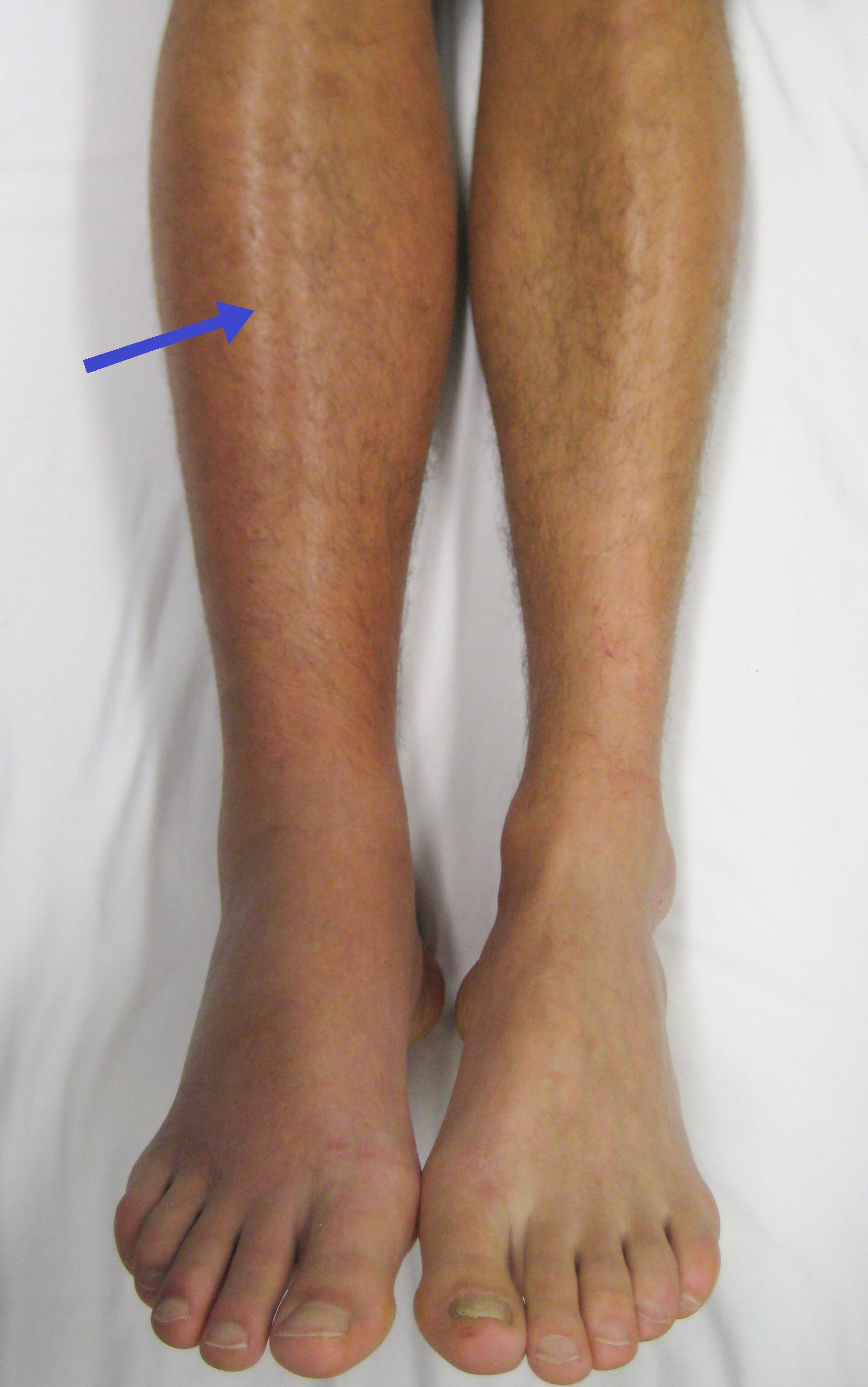
- DVT: unilateral leg swelling, warmth, tenderness (Homan sign unreliable)
- Massive PE: syncope, hypotension, JVD, loud P2, RV heave
Vignette: A 58-year-old woman has acute dyspnea and pleuritic chest pain 5 days after total knee replacement. HR 112, RR 24, SpO₂ 90%, lungs clear, BP 128/78 (stable).
Approach — patient is hemodynamically stable, so risk-stratify:
- Wells score. PE likely (>4) → go straight to CTPA (CT pulmonary angiography, the test of choice).
- PE unlikely (≤4) → D-dimer; negative rules out (high NPV), positive → CTPA.
- PERC rule can exclude PE in a patient already judged low-risk by gestalt — without even a D-dimer.
Here: PE is the leading diagnosis + recent surgery + tachycardia push Wells >4 → next best step = CTPA, not D-dimer (which would be non-specifically elevated post-op anyway).
If contrast is contraindicated (renal failure, contrast allergy) → V/Q scan.
- CTPA = confirmatory test of choice → intraluminal filling defect
- Compression ultrasound = test of choice for DVT
- D-dimer: very sensitive, poor specificity → only useful to rule OUT at low/intermediate pretest probability
- ABG: hypoxemia, hypocapnia, respiratory alkalosis, widened A–a gradient
- ECG: sinus tachycardia = most common; S1Q3T3 and new RBBB = RV strain (classic but uncommon)
- CXR: usually normal; may show Hampton hump (wedge-shaped infarct), Westermark sign (focal oligemia), Fleischner sign (enlarged central pulmonary artery)
- Troponin/BNP ↑ with RV dysfunction on echo (McConnell sign — RV free-wall hypokinesis with apical sparing) = submassive PE, worse prognosis
- PFTs are not used for acute PE; chronic thromboembolic pulmonary hypertension (CTEPH) shows a reduced DLCO

Risk Stratification Drives Treatment
| Category | Hemodynamics | RV strain (troponin/BNP/echo) | Management |
|---|---|---|---|
| Low-risk | Normal BP | Absent | Anticoagulation; consider outpatient if PESI low |
| Submassive (intermediate) | Normotensive | Present | Anticoagulate + admit/monitor; thrombolysis case-by-case |
| Massive (high-risk) | SBP <90 / shock | Present | Thrombolysis (tPA); if contraindicated → catheter-directed therapy or embolectomy |
- Virchow's triad (why clots form): Stasis · Hypercoagulability · Endothelial injury
- S1Q3T3 (acute cor pulmonale on ECG): deep S in lead I, Q wave + inverted T in lead III = RV strain — classic, though sinus tachycardia is commoner
- Wells score → PE likely vs unlikely decides CTPA vs D-dimer
- Homan sign (calf pain on dorsiflexion): classically taught for DVT but insensitive and nonspecific — don't rely on it
Vignette: A 66-year-old man collapses hours after a 14-hour flight. BP 78/40, HR 130, JVD, SpO₂ 84%; bedside echo shows a dilated, hypokinetic RV (McConnell sign).
This is massive (high-risk) PE with obstructive shock. Next best step:
- Resuscitate: O₂, cautious IV fluids (aggressive volume worsens RV distension), vasopressors; start heparin.
- Systemic thrombolysis (alteplase/tPA) — the answer for hemodynamically unstable PE with no contraindication.
- If thrombolysis is contraindicated (active bleeding, recent stroke/surgery) or fails → catheter-directed thrombolysis or surgical embolectomy.
If the patient is too unstable to travel, treat empirically — don't wait for CTPA. Place an IVC filter only when anticoagulation is contraindicated or PE recurs despite adequate anticoagulation.
Anticoagulation: Picking the Agent
| Scenario | Preferred agent | Key point |
|---|---|---|
| Most stable patients | DOAC (apixaban, rivaroxaban) | No routine monitoring; these two need no heparin bridge (unlike dabigatran/edoxaban) |
| Cancer-associated | DOAC or LMWH | LMWH long the standard; caution with GI/GU tumors on DOAC (bleeding) |
| Pregnancy | LMWH (enoxaparin) | Warfarin & DOACs contraindicated |
| Warfarin therapy | Warfarin + heparin bridge | Overlap ≥5 d until INR 2–3; bridge covers transient protein C drop |
| HIT | Argatroban / bivalirudin | Stop all heparin; avoid warfarin until platelets recover |
- Provoked by a transient/reversible factor (e.g., surgery) → 3 months
- Unprovoked → at least 3 months, then reassess for extended/indefinite therapy
- Recurrent VTE or active cancer → indefinite anticoagulation
- IVC filter: anticoagulation contraindicated (active bleeding) or recurrent PE despite therapeutic anticoagulation
- Board look-alikes: fat embolism (long-bone fracture → dyspnea + petechiae + confusion), amniotic fluid embolism (labor → DIC + shock), air embolism
- Prevention in hospitalized/post-op patients: early ambulation, LMWH prophylaxis, pneumatic compression
Practice Pulmonary now
Board-style questions, spaced-repetition flashcards, and a Socratic AI tutor — free to start.