Psoriasis & Papulosquamous Disorders
Board-focused review of psoriasis and the papulosquamous differential (lichen planus, pityriasis rosea, pityriasis rubra pilaris, secondary syphilis): classic morphology, bedside signs, histology/DIF buzzwords, high-yield associations, and the stepwise treatment ladder — anchored by the two exam traps (avoid systemic steroids in plaque psoriasis; rule out syphilis when palms/soles are involved).
Framing: what "papulosquamous" means and why psoriasis is the anchor
Papulosquamous disorders share one primary morphology: papules and plaques topped by scale. The board prototype is psoriasis, and most questions test how to separate it from its mimics — lichen planus, pityriasis rosea, pityriasis rubra pilaris, and secondary syphilis.
Psoriasis vulgaris affects ~2–3% of people, with a bimodal onset (teens–20s, then 50s–60s) and a strong genetic basis (HLA-Cw6 / PSORS1). It is immune-mediated, not simply "skin that grows too fast": activated dermal dendritic cells release IL-23, driving Th17 cells to secrete IL-17A and IL-22, while TNF-α amplifies the loop. The downstream result is keratinocyte hyperproliferation with markedly shortened epidermal transit (~4 days vs the normal ~28) — which is why nuclei are retained in the cornified layer (parakeratosis). This IL-23/IL-17 axis is the direct target of modern biologics.
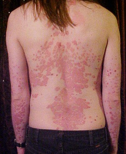
- Classic lesion: sharply demarcated salmon-pink plaques with silvery (micaceous) scale on extensor surfaces (elbows, knees), scalp, and gluteal cleft — umbilicus often involved.
- Bedside signs: Auspitz sign (pinpoint bleeding when scale is lifted, from dilated papillary capillaries under a thinned suprapapillary plate) and Koebner phenomenon (lesions along lines of trauma).
- Histology buzzwords: Munro microabscesses (neutrophils in the stratum corneum), spongiform pustules of Kogoj (neutrophils in the stratum spinosum), confluent parakeratosis with loss of the granular layer, regular (even) acanthosis with elongated rete ridges, and dilated papillary capillaries.
- Guttate psoriasis: abrupt crop of small "raindrop" plaques in a child/young adult ~1–3 weeks after streptococcal pharyngitis — check throat culture/ASO.
- Nails: pitting, "oil-drop" (salmon) sign, onycholysis, subungual hyperkeratosis.
- Psoriatic arthritis: seronegative spondyloarthropathy with DIP disease, dactylitis (sausage digit), and "pencil-in-cup" deformity on X-ray; axial disease tracks with HLA-B27.
- Do NOT use systemic corticosteroids for plaque psoriasis — taper/withdrawal can trigger a generalized pustular (von Zumbusch) flare or erythroderma.
- Lichen planus contrast (the classic distractor): orthokeratosis with NO parakeratosis, wedge-shaped hypergranulosis, sawtooth rete ridges, a band-like lymphocytic infiltrate at the dermoepidermal junction, and Civatte (colloid) bodies; DIF = shaggy fibrinogen at the basement membrane. Strong association with hepatitis C.
- Chronic plaque (vulgaris) — most common; symmetric extensor plaques, scalp, and intergluteal cleft.
- Guttate — post-streptococcal "raindrop" papules on the trunk; frequently self-limited.
- Inverse (flexural) — shiny, sharply marginated red plaques with little or no scale in axillae, inframammary, and inguinal folds (moisture removes scale); mimics candidal intertrigo.
- Pustular — localized palmoplantar pustulosis, or generalized (von Zumbusch) with sheets of sterile pustules, fever, and leukocytosis — a dermatologic emergency.
- Erythrodermic — >90% BSA erythema and scale with impaired thermoregulation and fluid/protein loss — also an emergency.
Provoking factors to know: streptococcal infection (guttate), trauma (Koebner), drugs — β-blockers, lithium, antimalarials, interferon, and systemic-steroid withdrawal, and HIV (can be severe/explosive).
Comorbidities (frequently tested): metabolic syndrome, cardiovascular disease, obesity, depression, uveitis, and inflammatory bowel disease. Treat psoriasis as a systemic inflammatory condition, not a skin-only problem.

One-look differential of the papulosquamous disorders
| Disorder | Morphology & distribution | Histology / DIF buzzword | Key association / next step |
|---|---|---|---|
| Psoriasis | Well-demarcated salmon plaques, silvery scale, extensor surfaces, scalp, gluteal cleft; Auspitz + Koebner | Munro microabscess, Kogoj pustule, parakeratosis + absent granular layer, even acanthosis | Strep → guttate; HLA-Cw6 / HLA-B27; avoid systemic steroids |
| Lichen planus | Violaceous, flat-topped, polygonal pruritic papules; wrists/ankles/flexors; Wickham striae; oral lacy white plaques | Sawtooth rete, wedge hypergranulosis, band-like infiltrate, Civatte bodies; DIF shaggy fibrinogen | Hepatitis C; drug-induced (thiazides, ACE-i, antimalarials, gold) |
| Pityriasis rosea | Herald patch → oval plaques with collarette scale along cleavage lines ("Christmas-tree" on back) | Superficial perivascular lymphocytes, focal parakeratosis, extravasated RBCs (nonspecific) | HHV-6/7; self-limited 6–8 wk; if palms/soles involved → RPR to exclude syphilis |
| Pityriasis rubra pilaris | Orange-red plaques, follicular hyperkeratotic papules, islands of sparing, waxy palmoplantar keratoderma | Alternating ortho/parakeratosis ("checkerboard"), follicular plugging | Cephalocaudal spread; usually idiopathic |
| Secondary syphilis | Coppery papulosquamous eruption involving palms and soles; condyloma lata; "moth-eaten" alopecia | Plasma-cell–rich infiltrate; spirochetes on immunostain | RPR/VDRL, then treponemal test; treat with penicillin |
The two interface patterns you must separate: psoriasis vs lichen planus
Psoriasis is a "neutrophilic, proliferative" pattern. Rapid turnover yields confluent parakeratosis and a diminished/absent granular layer; neutrophils migrate upward to form Munro microabscesses in the stratum corneum and spongiform pustules of Kogoj in the spinous layer. Rete ridges are regularly (evenly) elongated with thinned suprapapillary plates overlying dilated, tortuous papillary capillaries — the anatomic basis of the Auspitz sign.
Lichen planus is a "lymphocytic, interface" pattern. There is compact orthohyperkeratosis with NO parakeratosis, wedge-shaped hypergranulosis, and a dense band-like (lichenoid) lymphocytic infiltrate hugging the dermoepidermal junction. That infiltrate damages the basal layer (vacuolar/interface degeneration), producing Civatte / colloid bodies (apoptotic keratinocytes) and sawtooth rete ridges. DIF shows shaggy fibrinogen deposition along the basement membrane zone.
One-line discriminator: retained nuclei in the cornified layer (parakeratosis) = psoriasis; a thickened granular layer with no parakeratosis = lichen planus.

Lichen planus — the "6 P's": Pruritic, Purple (violaceous), Polygonal, Planar (flat-topped), Papules / Plaques. Add the seventh clue at the bedside: Wickham striae — fine white reticulated lines across the surface.
Psoriasis neutrophil collections — keep the layers straight: Munro microabscess sits in the stratum cor·neu·M (both carry the "m" — the more superficial layer); the Kogoj spongiform pustule sits deeper, in the spinous layer.
(These are the conventional teaching mnemonics — no fabricated associations added.)
Limited disease (roughly <5–10% BSA):
- First-line: topical corticosteroids, usually combined with a vitamin D3 analogue (calcipotriene/calcipotriol).
- Face and flexures: topical calcineurin inhibitors (tacrolimus/pimecrolimus) to avoid steroid atrophy.
- Adjuncts: tazarotene, coal tar, keratolytics (salicylic acid); medicated scalp solutions/foams.
Moderate-to-severe disease (BSA >5–10%, or disabling/special-site):
- Phototherapy: narrow-band UVB is first-line non-systemic therapy.
- Conventional systemics: methotrexate (also treats psoriatic arthritis), cyclosporine (fast rescue for acute flares), acitretin (best for pustular/erythrodermic disease; teratogenic — avoid pregnancy for 3 years), apremilast (PDE-4 inhibitor).
- Biologics (screen for latent TB and hepatitis first): TNF-α inhibitors (adalimumab, etanercept, infliximab), IL-12/23 (ustekinumab), IL-17 (secukinumab, ixekizumab, brodalumab), IL-23 (guselkumab, risankizumab). IL-17 inhibitors can unmask or worsen IBD — prefer IL-23 blockade or ustekinumab if Crohn's coexists.
Guttate: often self-limited; treat the streptococcal trigger and add NB-UVB if widespread.
Never attempt to control plaque psoriasis with systemic corticosteroids.
Pityriasis rosea and its can't-miss mimic
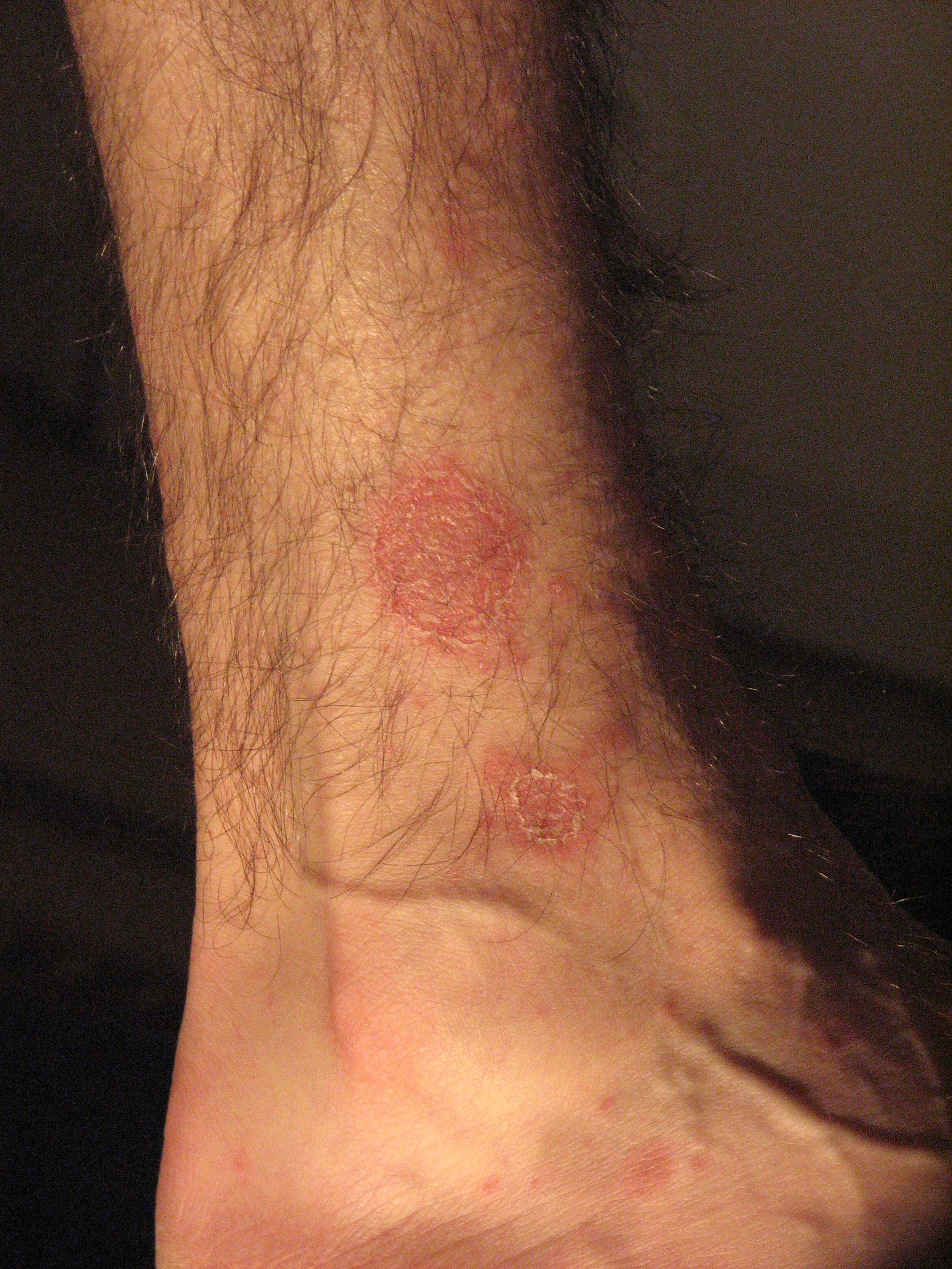
Pityriasis rosea begins with a solitary herald patch (a 2–5 cm oval plaque with a trailing collarette of scale), followed days later by a generalized eruption of smaller oval plaques oriented along skin cleavage lines, producing the "Christmas-tree" pattern across the back. It is linked to HHV-6/HHV-7 reactivation and resolves spontaneously in 6–8 weeks; management is symptomatic (emollients, mid-potency topical steroids, antihistamines).
The board trap: secondary syphilis can look nearly identical but characteristically involves the palms and soles and travels with generalized lymphadenopathy, condyloma lata, mucous patches, and "moth-eaten" alopecia. In any sexually active patient — or whenever palms/soles are involved — order an RPR/VDRL before committing to pityriasis rosea. Also on the differential: guttate psoriasis (smaller, more silvery, scalier papules, often post-strep) and a lichenoid/pityriasiform drug eruption (e.g., from ACE inhibitors or gold).
Practice Dermatology now
Board-style questions, spaced-repetition flashcards, and a Socratic AI tutor — free to start.