Primary Immunodeficiencies
A board-focused map of primary immunodeficiencies organized by the four arms of host defense — B cell, T cell, phagocyte, and complement — linking each defective molecule to its signature infection pattern and syndrome. Includes comparison tables, defect→disease vignettes, and the classic USMLE mnemonics.
The Framework: Four Arms, Four Infection Patterns
Primary immunodeficiencies (PIDs) are inherited defects in one of four arms of host defense: B cells (antibody/humoral), T cells (cellular), phagocytes, and complement. Step 1 vignettes almost always hand you three clues — the age at onset, the class of organism, and a syndromic feature — and ask you to name the broken molecule.
- Antibody (B-cell) defects present after ~6 months, once maternal IgG wanes → recurrent encapsulated bacterial (S. pneumoniae, H. influenzae), sinopulmonary, and GI infections (Giardia), plus enteroviruses.
- T-cell / combined defects present in the first months of life → viral, fungal (Candida), and opportunistic (PCP) infections with failure to thrive.
- Phagocyte defects → catalase-positive organisms and fungi (CGD), poor wound healing, and recurrent skin/soft-tissue abscesses; the adhesion subset (LAD) adds pus-less lesions and delayed umbilical cord separation.
- Complement defects → Neisseria (terminal C5–C9) or recurrent pyogenic/autoimmune disease (early components).
Master the logic organism + timing + syndrome → defect, and the reverse (defect → expected infection), because the exam runs it both ways.
- Selective IgA deficiency = most common PID; usually asymptomatic; classic trap is anaphylaxis to IVIG/blood products from anti-IgA antibodies.
- X-linked (Bruton) agammaglobulinemia = BTK defect → B cells can't mature → absent B cells, scant tonsils/lymph nodes, all Ig classes low; boys, after 6 months.
- SCID: most common cause is X-linked IL-2Rγ (common γ-chain) mutation; ADA deficiency is autosomal recessive; absent thymic shadow, ↓ TRECs on newborn screen; no live vaccines; cure = stem cell transplant.
- DiGeorge = 22q11.2 deletion, failed 3rd/4th pharyngeal pouches → no thymus/parathyroids → ↓ T cells, hypocalcemic tetany, conotruncal cardiac defects.
- Hyper-IgM = usually CD40L (CD40LG) defect on T cells → no class switching → ↑ IgM, ↓ IgG/IgA/IgE; watch for Pneumocystis and Cryptosporidium.
- Chronic granulomatous disease = NADPH oxidase defect → no respiratory burst → catalase-positive organisms; diagnose with dihydrorhodamine (DHR) flow or nitroblue tetrazolium.
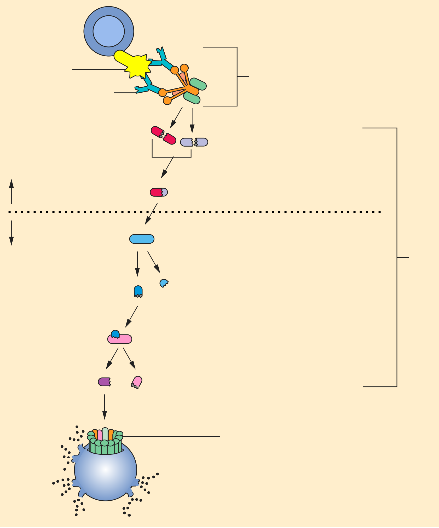
- Terminal complement (C5–C9 / MAC) deficiency → recurrent Neisseria; C1 esterase inhibitor deficiency → hereditary angioedema (↑ bradykinin, ↓ C4), and ACE inhibitors are contraindicated.
B-Cell (Humoral) Deficiencies
| Disease | Defect (gene/molecule) | Mechanism | Key clinical clues |
|---|---|---|---|
| Selective IgA deficiency | ↓ IgA (mucosal B-cell maturation; cause often unknown) | Low serum IgA, normal IgG/IgM | Most common PID; usually asymptomatic; sinopulmonary + GI infections, Giardia, atopy/autoimmunity; anaphylaxis to blood products / IVIG (anti-IgA Abs) |
| X-linked (Bruton) agammaglobulinemia | BTK (Bruton tyrosine kinase); X-linked recessive | B cells arrest at pre-B stage → absent mature B cells & plasma cells, all Ig low | Boys after 6 mo; recurrent encapsulated bacteria, enterovirus (echovirus) meningoencephalitis, Giardia; absent tonsils/lymph nodes; no live vaccines |
| Common variable immunodeficiency (CVID) | Defective B-cell differentiation (heterogeneous) | Normal B-cell number but ↓ plasma cells, ↓ IgG ± IgA/IgM | Onset teens–30s; recurrent sinopulmonary infections, bronchiectasis; ↑ autoimmune disease and lymphoma |

T-Cell and Combined (B + T) Deficiencies
| Disease | Defect (gene/molecule) | Mechanism | Key clinical clues |
|---|---|---|---|
| Thymic aplasia (DiGeorge) | 22q11.2 microdeletion; 3rd/4th pharyngeal pouch failure | Absent thymus & parathyroids → ↓ T cells, ↓ PTH | Tetany (hypocalcemia), conotruncal heart defects (tetralogy, truncus), abnormal facies, cleft palate; viral/fungal infections |
| IL-12 receptor deficiency | IL-12 receptor; autosomal recessive | ↓ Th1 response → ↓ IFN-γ | Disseminated mycobacteria (incl. after BCG) and endemic fungi |
| Autosomal dominant Hyper-IgE (Job) | STAT3 (loss of function) | ↓ Th17 → impaired neutrophil recruitment | Coarse Facies, cold (noninflamed) staph Abscesses, retained primary Teeth, ↑IgE, Dermatitis/eczema; eosinophilia, bone fractures |
| SCID | IL-2Rγ (common γ-chain) X-linked (most common); ADA deficiency (AR); RAG1/2 | No functional T cells ± B/NK; RAG → failed VDJ recombination | Early severe viral/fungal/bacterial + PCP, chronic diarrhea, thrush, FTT; absent thymic shadow, ↓ TRECs; treat with transplant |
| Hyper-IgM syndrome | CD40L (CD40LG) on Th cells (X-linked); or AID | No class switching → ↑ IgM, ↓ IgG/IgA/IgE | Severe pyogenic infections + opportunists: Pneumocystis, Cryptosporidium, CMV |
| Wiskott-Aldrich | WAS gene (WASp); X-linked recessive | Leukocytes/platelets can't reorganize actin cytoskeleton | Thrombocytopenia (small platelets), Eczema, Recurrent infections; ↑ IgE/IgA, ↓ IgM; ↑ autoimmunity & malignancy |
| Ataxia-telangiectasia | ATM (dsDNA break repair); autosomal recessive | Defective cell-cycle checkpoint/DNA repair; ↓ IgA | Cerebellar ataxia, telangiectasias, ↑ AFP, radiosensitivity, ↑ lymphoma/leukemia |
Phagocyte and Complement Defects
| Disease | Defect (gene/molecule) | Mechanism | Key clinical clues |
|---|---|---|---|
| Leukocyte adhesion deficiency type 1 | CD18 / β2-integrin (LFA-1); autosomal recessive | Phagocytes can't adhere/extravasate to sites of infection | Recurrent skin/mucosal bacterial infections without pus, delayed umbilical cord separation (>30 d), poor wound healing, ↑ circulating neutrophils |
| Chédiak-Higashi | LYST (lysosomal trafficking regulator); AR | Failed phagolysosome fusion; microtubule dysfunction | Recurrent pyogenic (staph/strep), partial albinism, peripheral neuropathy, giant granules in leukocytes, pancytopenia; accelerated (HLH) phase |
| Chronic granulomatous disease (CGD) | NADPH oxidase (often gp91phox, X-linked) | No respiratory burst / superoxide → can't kill catalase(+) organisms | Catalase-positive infections (S. aureus, Serratia, Nocardia, Burkholderia, Aspergillus); granulomas; dx = DHR flow / nitroblue tetrazolium |
| C1 esterase inhibitor deficiency | SERPING1 (C1-INH); autosomal dominant | Unregulated bradykinin (and complement) generation | Hereditary angioedema (recurrent, nonpitting, nonpruritic/non-urticarial); ↓ C4; ACE inhibitors contraindicated |
| C3 deficiency | C3 | Loss of central opsonin & pathway convergence | Severe recurrent pyogenic (encapsulated) infections; type III hypersensitivity |
| Terminal complement (C5–C9) | C5–C9 / MAC | Cannot form membrane attack complex | Recurrent Neisseria (meningococcal, gonococcal); ↓ CH50 |
- A 9-month-old boy has had recurrent otitis media, two pneumonias, and one episode of echovirus meningoencephalitis. Exam shows no palpable tonsils. Flow cytometry: absent CD19+ B cells; all Ig classes low. → BTK defect = X-linked (Bruton) agammaglobulinemia.
- A neonate seizes from hypocalcemia and has a murmur of truncus arteriosus; CXR shows an absent thymic shadow; FISH reveals a 22q11.2 deletion. → DiGeorge (thymic + parathyroid aplasia).
- A 2-year-old boy with eczema, petechiae from thrombocytopenia (small platelets), and recurrent encapsulated infections. → WAS gene = Wiskott-Aldrich.
- A 6-year-old boy with recurrent S. aureus and Serratia abscesses and hepatic granulomas; dihydrorhodamine flow shows no oxidation (no green fluorescence). → NADPH oxidase defect = chronic granulomatous disease.
- A college freshman presents with a second episode of meningococcemia; CH50 is low. → Terminal complement (C5–C9 / MAC) deficiency.
- A 10-month-old with chronic diarrhea, thrush, PCP, and failure to thrive; newborn screen had shown low TRECs. → SCID (X-linked IL-2Rγ or ADA deficiency).
- CATCH-22 → DiGeorge: Cardiac (conotruncal) defects, Abnormal facies, Thymic aplasia, Cleft palate, Hypocalcemia — chromosome 22q11.2.
- FATED → AD Hyper-IgE (Job): coarse Facies, cold staph Abscesses, retained primary Teeth, ↑IgE, Dermatitis (eczema).
- WATER → Wiskott-Aldrich: Wiskott-Aldrich, Thrombocytopenia (small platelets), Eczema, Recurrent infections.
- Job syndrome — biblical Job was "smote with boils" = the cold staph skin abscesses.
- Catalase-positive organisms (CGD) — "Cats Need PLACESS": Nocardia, Pseudomonas, Listeria, Aspergillus, Candida, E. coli, Staph aureus, Serratia (± Burkholderia cepacia).
- Encapsulated bacteria (S. pneumoniae, Hib, N. meningitidis) → antibody, complement, or splenic defect.
- Recurrent Neisseria → terminal complement (C5–C9 / MAC) deficiency.
- Catalase-positive bacteria/fungi (S. aureus, Serratia, Nocardia, Burkholderia, Aspergillus) → phagocyte oxidative burst (CGD).
- Candida / PCP / CMV, disseminated viral infection + FTT in infancy → T-cell / combined defect (SCID, DiGeorge).
- Disseminated mycobacteria (including after BCG) → IL-12 / IFN-γ axis (IL-12R deficiency).
- Giardia, enterovirus (echovirus), recurrent sinopulmonary → humoral defect (Bruton, IgA deficiency, CVID).
- No pus + delayed umbilical cord separation → leukocyte adhesion deficiency (CD18).
Practice Immunology now
Board-style questions, spaced-repetition flashcards, and a Socratic AI tutor — free to start.