Potassium Disorders (Hypo/Hyperkalemia)
A Step 2 CK-focused breakdown of hypo- and hyperkalemia covering pathophysiology (internal vs external balance), classic causes and ECG buzzwords, lab/urine localization, and the emergency next-best-step management ladders — stabilize → shift → eliminate for hyperkalemia, and replace K+ plus Mg for hypokalemia. Built around vignette buzzwords (U waves, peaked T waves) and the digoxin links boards love to test.
Overview: Potassium Balance
Potassium is the major intracellular cation (~98% inside cells). The steep transmembrane gradient, maintained by the Na+/K+-ATPase, sets the resting membrane potential, so small serum changes produce large effects on excitable tissue — heart, nerve, and muscle. Balance has two arms:
- Internal (ICF↔ECF shifts): insulin, β2-agonists, and alkalosis drive K+ into cells (lowers serum K+); acidosis (especially mineral/non-anion-gap acids), insulin deficiency, cell lysis, and digoxin (Na/K-ATPase block) push K+ out (raises serum K+).
- External (excretion): the distal nephron (principal cells, aldosterone-driven) is the main route; GI (colonic) loss is minor except in advanced CKD, where it becomes a meaningful compensatory route.
Because of shifts, serum K+ is a poor mirror of total-body stores — always interpret it alongside acid–base status, renal function, and the ECG. Both extremes are lethal via arrhythmia, which is why potassium is one of the highest-yield electrolyte topics on Step 2 CK.
- Threshold: <3.5 mEq/L; <2.5 is severe/emergent.
- GI loss: diarrhea (with non-anion-gap metabolic acidosis). Vomiting/NG suction lose K+ indirectly — volume depletion → aldosterone + bicarbonaturia drive renal K+ wasting (metabolic alkalosis).
- Renal loss: loop/thiazide diuretics; primary hyperaldosteronism (Conn: HTN + hypokalemia + alkalosis); Cushing; licorice/apparent mineralocorticoid excess; Bartter (loop-like, normal BP); Gitelman (thiazide-like: hypocalciuria + hypomagnesemia); Liddle (low renin + low aldo, HTN); RTA types 1 & 2.
- Transcellular shift: insulin, β-agonists, alkalosis, refeeding, hypokalemic periodic paralysis, high turnover (e.g., B12-treated megaloblastic anemia).
- ECG: flat/inverted T waves, ST depression, prominent U waves, long QT → torsades; potentiates digoxin toxicity.
- Localize: spot urine K+ (or TTKG) — low = extrarenal/GI loss; inappropriately high = renal wasting.
- Pearl: repletion-refractory hypokalemia → check and replace magnesium.
- Threshold: >5.0–5.5 mEq/L; >6.5 or ECG changes = emergency.
- Pseudohyperkalemia: hemolyzed sample, fist-clenching/tourniquet, marked thrombocytosis/leukocytosis — repeat before treating if the patient is well with a normal ECG.
- ↓ Excretion: AKI/CKD (esp. oliguric); type 4 RTA / hyporeninemic hypoaldosteronism (diabetic); Addison; drugs — ACEi/ARB, K-sparing diuretics (spironolactone, amiloride), NSAIDs, trimethoprim, heparin, calcineurin inhibitors.
- Shift out of cells: metabolic acidosis, insulin deficiency (DKA — driven mainly by insulin lack + hyperosmolality), nonselective β-blockers, digoxin toxicity, cell lysis (rhabdomyolysis, tumor lysis, hemolysis), succinylcholine, hyperkalemic periodic paralysis.
- ECG progression: peaked T waves (earliest) → PR prolongation → loss of P waves → wide QRS → sine wave → VF/asystole.
- Classic combos: crush injury/CKD; TMP-SMX in a transplant patient; ACEi + spironolactone + NSAID in CKD.

Hypo- vs Hyperkalemia at a Glance
| Feature | Hypokalemia (<3.5) | Hyperkalemia (>5.0–5.5) |
|---|---|---|
| Common causes | Diuretics, vomiting/diarrhea, hyperaldosteronism, ↓Mg | Renal failure, ACEi/ARB + K-sparing drug, acidosis, cell lysis |
| Buzzwords | U waves, licorice, bulimia | Peaked T waves, crush injury, TMP-SMX |
| ECG | Flat T, ST depression, U waves, long QT → torsades | Peaked T → loss of P → wide QRS → sine wave → VF |
| Acid–base | Often metabolic alkalosis | Often metabolic acidosis |
| First emergent step | ECG; KCl (oral/IV) + replace Mg | ECG; IV calcium (membrane stabilization) |
| Digoxin link | Worsens dig toxicity | Hyperkalemia = marker of severe dig toxicity → give DigiFab |
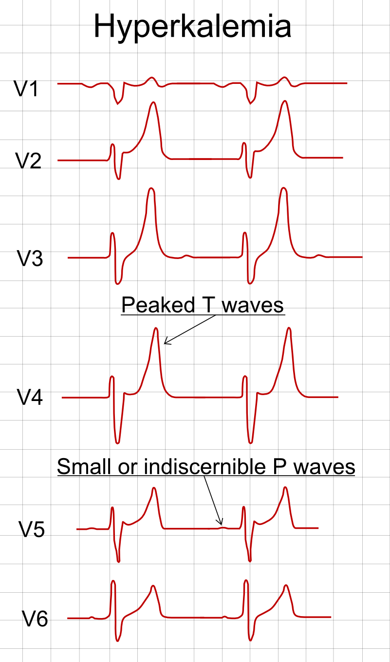
Stem: A 68-year-old man with CKD stage 4 on lisinopril and spironolactone presents with generalized weakness and palpitations. K+ 7.3 mEq/L, HCO3 18. ECG: peaked T waves with a widening QRS.
Diagnosis: Life-threatening hyperkalemia with ECG changes (drug-induced ↓ excretion on a CKD background).
Next best step (in order):
- IV calcium gluconate immediately — stabilizes the myocardial membrane, works within minutes (does not lower K+); repeat if ECG changes persist.
- Shift K+ into cells: regular insulin + dextrose (± nebulized albuterol; add bicarbonate only if acidemic).
- Remove K+: loop diuretic (if making urine), GI binder (sodium zirconium cyclosilicate or patiromer), or hemodialysis — definitive, especially if anuric/refractory.
- Stop the ACEi and spironolactone.
Trap: insulin/calcium/albuterol are temporizing — you must still remove K+ from the body.
Stem: A 34-year-old woman with chronic alcohol use and poor intake has K+ 2.8 despite receiving 80 mEq of IV KCl. She reports cramps; Mg 1.2 mg/dL (low), and the ECG shows a prolonged QT.
Diagnosis: Hypokalemia refractory to repletion because of coexisting hypomagnesemia. Low intracellular Mg2+ relieves its normal block on the ROMK channel, so the distal nephron keeps secreting (wasting) K+ — serum K+ will not correct until Mg is replaced.
Next step:
- Replace magnesium (IV MgSO4) alongside potassium — this is the key testable move.
- Continue KCl repletion; recheck K+ and Mg.
- Monitor the ECG for torsades given the long QT.
- Address the cause: nutrition, thiamine before glucose, and refeeding precautions.
The three-step framework boards actually test — remember the categories in order, not a shaky acronym:
- Stabilize the myocardium: IV calcium (gluconate peripherally; chloride only via central line) — give first if ECG changes; onset in minutes, effect is short-lived, and it does not lower serum K+.
- Shift K+ into cells: insulin + glucose (onset ~15–30 min, the workhorse); nebulized albuterol (additive); sodium bicarbonate only if acidemic (weak/adjunctive, minimal benefit in ESRD).
- Eliminate K+ from the body: loop diuretic (if still making urine); GI cation binders — patiromer or sodium zirconium cyclosilicate (older SPS/"Kayexalate" is slow and carries a colonic-necrosis risk, so it's fallen out of favor for acute use); hemodialysis — definitive, especially if anuric or refractory.
Key point: only calcium protects the heart electrically; only diuretics, binders, and dialysis actually remove total-body K+ — shifting just buys time.
- Hyperkalemia order of action: (1) ECG/monitor, (2) stabilize (calcium), (3) shift (insulin+glucose, albuterol, bicarb if acidemic), (4) remove (loop diuretic, binder, dialysis).
- Calcium + digoxin: classic teaching warned against calcium in digoxin toxicity ("stone heart"); regardless, the exam answer for dig toxicity + hyperkalemia is digoxin-specific Fab (DigiFab).
- Hypokalemia: prefer oral KCl if tolerated; peripheral IV ≤10 mEq/hr (faster requires central access + continuous cardiac monitoring) — never bolus. Correct Mg first/simultaneously. Rule of thumb: a serum K+ fall of ~1 mEq/L reflects a 200–400 mEq total-body deficit.
- Always treat the cause: stop offending drugs; correct volume and acid–base disturbances.
- Avoid dextrose-alone shifting (no insulin) — a glucose load can transiently worsen hyperkalemia in insulin-deficient patients.
Practice Renal now
Board-style questions, spaced-repetition flashcards, and a Socratic AI tutor — free to start.