Pneumonia Types & Lung Abscess
A high-yield Step 1/Step 2 CK lesson on pneumonia (typical, atypical, aspiration) and lung abscess, threading pathophysiology to next-best-step management through classic vignette buzzwords. Emphasizes host/exposure-driven organism selection, empiric therapy, and cavitary aspiration disease.
Pathophysiology & Classification
Pneumonia is infection of the lung parenchyma, classified by setting — community-acquired (CAP), hospital-acquired (HAP, ≥48 h after admission), and ventilator-associated (VAP) — and by anatomic pattern. Lobar pneumonia fills an entire lobe with exudate (classically S. pneumoniae); bronchopneumonia is patchy and multifocal (Staph, gram-negatives); interstitial pneumonia is diffuse with a mononuclear/lymphocytic infiltrate (Mycoplasma, viruses). Legionella is grouped clinically as "atypical" but histologically drives a neutrophilic alveolar exudate and often produces patchy or lobar consolidation. Step 1 loves the four histologic phases of lobar pneumonia: congestion → red hepatization → gray hepatization → resolution. When aspirated anaerobes cause parenchymal necrosis and cavitation, the result is a lung abscess. Throughout, the host and exposure history drive the likely organism and therefore the next best step.

- S. pneumoniae — #1 CAP; rusty sputum, lobar, optochin-sensitive, bile-soluble, IgA protease; encapsulated → asplenia risk
- Mycoplasma — teens/young adults, dorms/military; dry cough, CXR worse than exam, cold agglutinins (IgM), bullous myringitis; no cell wall → macrolide/doxy, NOT β-lactams
- Klebsiella — alcoholics, aspiration; currant-jelly sputum, upper-lobe cavitation
- S. aureus — post-influenza, IVDU; necrotizing/cavitary
- Legionella — water/AC, cruises; high fever + relative bradycardia, diarrhea, hyponatremia, ↑LFTs; urine antigen (serogroup 1), buffered charcoal yeast extract agar
- Pseudomonas — CF, bronchiectasis, HAP/VAP, neutropenia
- Pneumocystis jirovecii — HIV CD4 <200; diffuse bilateral ground-glass, ↑LDH; silver stain; TMP-SMX (add steroids if PaO₂ <70 mmHg on room air or A–a gradient ≥35)
- Coxiella (Q fever, livestock/parturient animals), Chlamydophila psittaci (birds), H. influenzae (COPD)
Typical vs Atypical Pneumonia
| Feature | Typical | Atypical |
|---|---|---|
| Bugs | S. pneumoniae, H. flu, Klebsiella | Mycoplasma, Chlamydophila, Legionella, viruses |
| Onset | Abrupt, toxic | Gradual, "walking" |
| Sputum | Productive, purulent | Scant / dry |
| CXR | Lobar consolidation | Diffuse patchy / interstitial |
| WBC | Marked leukocytosis | Normal / mildly ↑ |
| First-line Rx | β-lactam (amoxicillin) | Macrolide / doxycycline |
Vignette: A 19-year-old college student has 2 weeks of malaise, low-grade fever, and a nagging dry cough. He looks well, yet CXR shows patchy bilateral infiltrates out of proportion to his mild symptoms. Cold agglutinins are positive.
Diagnosis: Atypical ("walking") pneumonia — *Mycoplasma pneumoniae*.
Next best step: Treat empirically with azithromycin (or doxycycline). Key trap: Mycoplasma has no cell wall, so β-lactams (amoxicillin, ceftriaxone) are useless. Boards-favorite complications: cold-agglutinin hemolytic anemia, erythema multiforme / SJS, and bullous myringitis.
Classic exam associations — when you see the phrase, name the organism:
- Rusty sputum → S. pneumoniae
- Currant-jelly sputum → Klebsiella
- Foul / putrid sputum → anaerobic lung abscess
- Cold agglutinins → Mycoplasma
- Air conditioner / water tower + hyponatremia → Legionella
- Post-influenza, cavitary → S. aureus
- Bird exposure → Chlamydophila psittaci; livestock / parturient animals → Coxiella (Q fever)
- Faget sign (fever with relative bradycardia) → Legionella
Lung Abscess — Aspiration & Anatomy
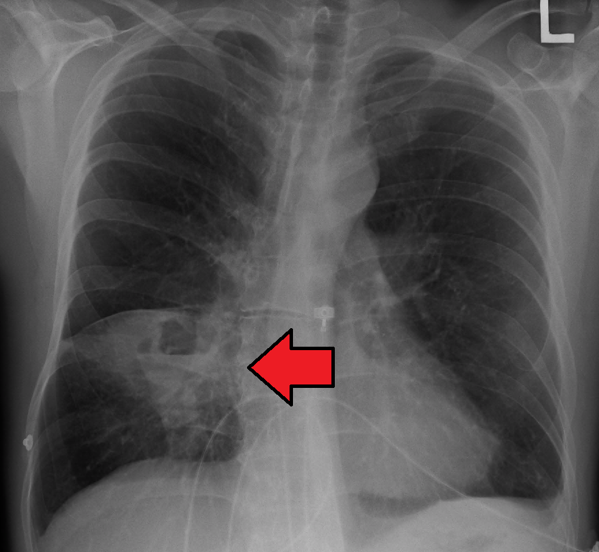
A lung abscess is a localized collection of pus with parenchymal necrosis and cavitation, most often from aspiration of oropharyngeal anaerobes (usually polymicrobial: Peptostreptococcus, Prevotella, Bacteroides, Fusobacterium). Risk factors impair airway protection: alcohol use, seizures, stroke, sedation, GERD/dysphagia, and poor dentition. The course is indolent over weeks — foul-smelling (putrid) sputum, night sweats, weight loss, low-grade fever — mimicking TB or malignancy. Aspirated material lands in dependent segments: supine → posterior segment of the RUL and superior segment of the RLL (the right main bronchus is wider and more vertical). Imaging shows a thick-walled cavity with an air-fluid level.
Vignette: A 55-year-old man with alcohol use disorder and poor dentition is brought in after a witnessed seizure. Over 3 weeks he developed cough productive of foul-smelling sputum, drenching night sweats, and 5-kg weight loss. CXR shows a cavity with an air-fluid level in the superior segment of the right lower lobe.
Diagnosis: Anaerobic lung abscess from aspiration.
Next best step: Start empiric ampicillin-sulbactam (β-lactam/β-lactamase inhibitor) — now preferred over clindamycin (C. diff risk) — the mainstay, given as prolonged therapy. Obtain a CT chest to characterize the cavity and exclude an obstructing mass (especially in older smokers). Percutaneous/surgical drainage is reserved for failure to respond.
- Dx of CAP: clinical features + infiltrate on CXR; PFTs have no diagnostic role. In severe/inpatient cases send sputum + blood cultures and Legionella + pneumococcal urine antigens.
- Severity — CURB-65: Confusion, Urea (BUN >19), RR ≥30, BP <90/60, age ≥65; score ≥2 favors admission.
- Outpatient, healthy: amoxicillin or doxycycline or a macrolide (IDSA 2019; macrolide monotherapy only where local pneumococcal resistance <25%).
- Outpatient + comorbidities: amox-clav/cephalosporin + macrolide/doxy, or a respiratory fluoroquinolone (levo/moxifloxacin).
- Inpatient (non-ICU): β-lactam + macrolide or respiratory FQ.
- HAP/VAP: empirically cover MRSA (vancomycin/linezolid) + Pseudomonas (piperacillin-tazobactam/cefepime).
- Older smoker: follow-up CXR to exclude post-obstructive pneumonia from malignancy.
Practice Pulmonary now
Board-style questions, spaced-repetition flashcards, and a Socratic AI tutor — free to start.