Pleural Effusion & Pneumothorax
A Step 2 CK-focused lesson on pleural effusion and pneumothorax, moving from pleural-space pathophysiology through bedside exam, imaging, and pleural-fluid analysis to next-best-step management, anchored by Light's criteria, drainage thresholds, and the tension-pneumothorax crash vignette. Includes two comparison tables, two clinical vignettes, and classic memory hooks.
Two ways the pleural space fails
The pleural space normally holds only ~10-20 mL of fluid, kept in balance by hydrostatic and oncotic (Starling) forces plus lymphatic drainage. The boards love to contrast the two ways this balance breaks: fluid accumulation (pleural effusion) versus air accumulation (pneumothorax).
Effusions split by mechanism. A transudate arises from altered hydrostatic/oncotic pressures across intact capillaries — heart failure (most common overall), cirrhosis, nephrotic syndrome. An exudate arises from increased capillary permeability or impaired lymphatic drainage from inflammation, infection, or cancer — pneumonia, malignancy, PE, TB, pancreatitis.
A pneumothorax is air in the pleural space that abolishes the negative intrapleural pressure holding the lung open, so the lung recoils and collapses. When a one-way valve lets air in but not out, intrathoracic pressure builds and shifts the mediastinum — a tension pneumothorax, which is obstructive shock and a clinical, not radiographic, diagnosis.
- Light's criteria — exudate if ANY one is met: pleural/serum protein > 0.5, pleural/serum LDH > 0.6, or pleural LDH > 2/3 the upper limit of normal serum LDH.
- Light's is very sensitive but mislabels some diuretic-treated CHF transudates as exudate; if suspected, a serum-pleural albumin gradient > 1.2 g/dL favors a transudate.
- Exam over fluid: dullness to percussion, decreased breath sounds, decreased tactile fremitus.
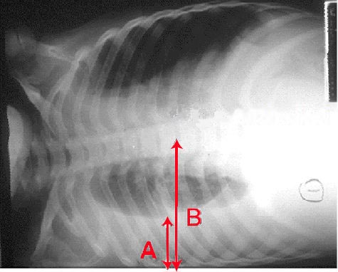
- CXR: blunting of the costophrenic angle + meniscus (needs ~200 mL on an upright PA film); a lateral decubitus film shows whether fluid is free-flowing (layers) vs loculated and detects small effusions (as little as ~5 mL); ultrasound best guides the tap and detects loculation.
- Thoracentesis for any new effusion of unclear cause — EXCEPT small bilateral effusions in clear-cut CHF, which you diurese first.
- Fluid clues: low glucose/low pH → empyema, TB, RA, malignancy; high amylase → pancreatitis or esophageal rupture; lymphocyte-predominant → TB/malignancy; milky, triglycerides > 110 mg/dL → chylothorax; grossly bloody → malignancy, trauma, PE.
Transudate vs exudate
| Feature | Transudate | Exudate |
|---|---|---|
| Mechanism | ↑ hydrostatic / ↓ oncotic, intact capillaries | ↑ permeability or ↓ lymphatic drainage |
| Light's criteria | Meets none | Meets ≥1 |
| Protein (pleural/serum) | ≤ 0.5 | > 0.5 |
| LDH (pleural/serum) | ≤ 0.6 | > 0.6 |
| Classic causes | CHF, cirrhosis, nephrotic syndrome | Pneumonia, malignancy, PE, TB, pancreatitis |
| Appearance | Clear, straw-colored | Cloudy, bloody, or purulent |
A 58-year-old man admitted for lobar pneumonia stays febrile on day 3 with worsening dyspnea. CXR shows a new moderate left effusion; a decubitus film shows it layers > 1 cm (free-flowing).
- Next best step: diagnostic thoracentesis with fluid analysis.
- Fluid returns pH 7.05, glucose 40 mg/dL, high LDH, positive Gram stain → complicated parapneumonic effusion / empyema.
- Management: chest tube (tube thoracostomy) drainage + continued antibiotics. Frank pus = empyema.
- Drainage triggers to memorize: pH < 7.2, glucose < 60 mg/dL, positive Gram stain/culture, loculation, or frank pus.
- Persistent loculated collections → intrapleural tPA + DNase; refractory → VATS / decortication.
Contrast: an uncomplicated parapneumonic effusion (pH > 7.2, no organisms) needs antibiotics alone — no chest tube.
- Primary spontaneous (PSP): tall, thin, young male smoker with no known lung disease; rupture of apical subpleural blebs. Classic association with connective-tissue disorders (Marfan, Ehlers-Danlos).
- Secondary spontaneous (SSP): underlying lung disease — COPD (most common), CF, TB, Pneumocystis (PCP) pneumonia in HIV, ILD, LAM, catamenial (thoracic endometriosis).
- Traumatic / iatrogenic: subclavian central line, thoracentesis or lung biopsy, barotrauma on mechanical ventilation, blunt/penetrating trauma.
- Presentation: sudden pleuritic chest pain + dyspnea; hyperresonance, decreased breath sounds, decreased tactile fremitus on the affected side.
- CXR: visceral pleural line with absent lung markings peripherally; deep sulcus sign on a supine film.
- Management (stable): small PSP, minimal symptoms → observation + supplemental O2 (high-flow O2 speeds pleural air resorption ~3-4×); large or symptomatic → needle aspiration or small-bore chest tube. SSP → chest tube + admit (poor reserve). Recurrence or persistent air leak → VATS bleb resection / pleurodesis.

A 24-year-old man on mechanical ventilation becomes acutely hypotensive and hypoxic. Exam: absent breath sounds and hyperresonance on the right, trachea deviated to the left, distended neck veins, BP 70/40.
- Diagnosis: tension pneumothorax — a clinical diagnosis.
- Next best step: immediate needle decompression — do NOT wait for a CXR.
- Needle: large-bore into the 2nd intercostal space, midclavicular line (or 5th ICS, anterior axillary line per current ATLS) on the affected side.
- Definitive treatment: chest tube (tube thoracostomy) after decompression.
Why shock: the one-way valve raises intrathoracic pressure, kinks the great veins, and drops venous return → obstructive shock. Hypotension + distended neck veins + absent unilateral breath sounds is the classic triad.
Effusion vs pneumothorax at the bedside
| Finding | Pleural effusion | Pneumothorax |
|---|---|---|
| Percussion | Dull | Hyperresonant |
| Breath sounds | Decreased | Decreased / absent |
| Tactile fremitus | Decreased | Decreased |
| Tracheal shift (large) | Away from side | Away (only if tension) |
| CXR | Blunted costophrenic angle, meniscus | Visceral pleural line, no distal markings |
| First move | Thoracentesis | Needle decompression (if tension) |
Light's criteria — exudate if ANY are met. Remember the cutoffs 0.5 / 0.6 / 2⁄3:
- Pleural/serum protein > 0.5
- Pleural/serum LDH > 0.6
- Pleural LDH > 2/3 the upper limit of normal serum LDH
Tracheal deviation direction:
- Pushed AWAY by added volume/pressure → tension pneumothorax and massive pleural effusion.
- Pulled TOWARD by lost volume → lobar collapse / atelectasis and pneumonectomy.
"Air and fluid push away; collapse pulls toward."
Practice Pulmonary now
Board-style questions, spaced-repetition flashcards, and a Socratic AI tutor — free to start.