Placenta, Twinning & Fetal Circulation
A high-yield Step 1 embryology lesson tying together placental structure, twin chorionicity/amnionicity by splitting time, and the fetal circulatory shunts with their adult remnants. Includes twin-twin transfusion and prostaglandin/ductus vignettes plus classic mnemonics.
The maternal–fetal interface, in one frame
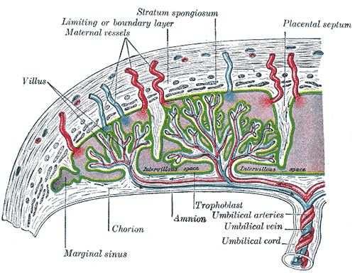
The placenta is the fetal–maternal exchange organ. Its fetal side (chorion frondosum + chorionic villi) interlocks with the maternal decidua basalis. Trophoblast differentiates into an inner cytotrophoblast ("cellular" stem-cell layer) and an outer syncytiotrophoblast, which erodes maternal spiral arteries, lacks MHC class I (immune privilege), and secretes hCG and human placental lactogen.
The umbilical cord carries two arteries (deoxygenated blood → placenta) and one vein (oxygenated blood → fetus), cushioned by Wharton jelly. Because the fetus oxygenates at the placenta rather than the lungs, three vascular shunts — ductus venosus, foramen ovale, ductus arteriosus — route blood past the liver and lungs, each leaving a named adult remnant.
Meanwhile, zygosity and the timing of embryo splitting set twin placentation. These three threads — placental structure, twinning, and fetal circulation — are among the densest, most testable embryology facts on Step 1.
- Syncytiotrophoblast secretes hCG → maintains the corpus luteum (progesterone) through the first trimester; detectable in blood ~1 wk, urine ~2 wks post-conception.
- Cytotrophoblast = inner, mitotically active stem-cell layer.
- Fetal component = chorion frondosum + villi; maternal component = decidua basalis.
- Umbilical cord = 2 arteries + 1 vein; the vein carries the oxygenated blood.
- Single umbilical artery → associated with congenital (cardiac, renal) and chromosomal anomalies.
- Velamentous insertion / vasa previa: unprotected fetal vessels cross the internal os → can rupture with the membranes → rapid fetal hemorrhage.
- Placenta accreta spectrum (accreta → increta → percreta): villi adhere to or invade the myometrium because of a defective decidua basalis; risk rises with prior C-section + placenta previa → severe postpartum hemorrhage.
Twin types by zygosity and splitting time
| Origin / MZ split timing | Chorion / Amnion | Key point |
|---|---|---|
| Dizygotic (2 eggs, 2 sperm) | Di / Di (always) | 2 placentas (may fuse); genetically like any siblings |
| Days 0–3 (before inner cell mass) | Di / Di (~⅓ of MZ) | 2 chorions, 2 amnions |
| Days 4–8 (blastocyst) | Mono / Di (most MZ) | shared placenta, separate sacs |
| Days 8–13 (after amnion forms) | Mono / Mono | one sac → cord-entanglement risk |
| >13 days (formed embryonic disc) | Conjoined | incomplete splitting |

Vignette: Ultrasound of monochorionic diamniotic twins shows one fetus with polyhydramnios, a distended bladder, and polycythemia, while the smaller co-twin has oligohydramnios, appears "stuck" against the uterine wall, and is anemic. Diagnosis, mechanism, and treatment?
Answer: Twin–twin transfusion syndrome (TTTS). Unbalanced arteriovenous anastomoses across the shared monochorionic placenta shunt blood from the donor (anemic, growth-restricted, oligohydramnios) to the recipient (polycythemic, volume-overloaded, polyhydramnios, risk of hydrops/heart failure).
- Requires monochorionic placentation — it cannot occur in dichorionic twins.
- Definitive treatment: fetoscopic laser photocoagulation of the connecting placental anastomoses.
- Umbilical vein carries the most oxygenated blood (~80% saturation) from placenta to fetus.
- Ductus venosus: umbilical vein → IVC, bypassing the liver.
- Foramen ovale: RA → LA — preferentially directs the most-oxygenated blood to the brain and heart via the ascending aorta.
- Ductus arteriosus: pulmonary artery → descending aorta, bypassing the fluid-filled, high-resistance lungs.
- The ductus arteriosus is held open by high PGE2 and low O2; at birth, ↑O2 + loss of placental prostaglandins → closure.
- Indomethacin (↓prostaglandins) closes a PDA; PGE1 (alprostadil) keeps it open for ductal-dependent lesions.

Fetal structure → adult remnant
| Fetal structure | Adult remnant |
|---|---|
| Umbilical vein | Ligamentum teres hepatis (round ligament, in the falciform ligament) |
| Ductus venosus | Ligamentum venosum |
| Ductus arteriosus | Ligamentum arteriosum |
| Foramen ovale | Fossa ovalis |
| Umbilical arteries (distal) | Medial umbilical ligaments (paired) |
| Allantois (urachus) | Median umbilical ligament (single, midline) |
| Notochord | Nucleus pulposus |
Vignette: A term newborn becomes progressively cyanotic within hours of birth. Echo shows the aorta arising from the right ventricle and the pulmonary artery from the left ventricle (transposition of the great arteries). Cyanosis worsens as the ductus arteriosus narrows. Next step?
Answer: Start prostaglandin E1 (alprostadil) to keep the ductus arteriosus patent, preserving the mixing/systemic flow the infant depends on until surgical correction (arterial switch).
Contrast: A preterm infant with a continuous "machinery" murmur from a patent ductus arteriosus is given indomethacin to close it.
This flips the fetal rule — after birth we normally want the ductus closed, except in ductal-dependent lesions, where we keep it open.
- Umbilical VEIN = oxygenated — "the Vein brings Vital O2 to the fetus." The two arteries carry deoxygenated blood away to the placenta (opposite of the systemic circulation).
- MediaN vs MediaL umbilical ligaments: MediaN = single midline = urachus (allantois remnant); MediaL (paired) = obLiterated umbiLical arteries.
- Ductus A → ligamentum arteriosum; ductus V → ligamentum venosum — the name barely changes, so the pairing is easy to recall.
Practice Embryology now
Board-style questions, spaced-repetition flashcards, and a Socratic AI tutor — free to start.