Pigmentation Disorders
A Step 1–focused dermatology lesson on pigmentation disorders, organized by mechanism (melanocyte loss vs. defective melanin synthesis vs. hormonal/UV-driven hyperpigmentation), with classic vignette buzzwords, comparison tables, and next-best-step decisions. Covers vitiligo, albinism, pityriasis versicolor, café-au-lait syndromes, melasma, Addison pigmentation, and the melanoma ABCDE rule.
Pathophysiology: melanin, melanocytes, and the framework
Melanocytes are neural-crest–derived dendritic cells in the stratum basale that make melanin inside melanosomes and transfer it to surrounding keratinocytes. Synthesis runs tyrosine → DOPA → dopaquinone → melanin, with the rate-limiting step catalyzed by tyrosinase (a copper-containing enzyme). Skin tone reflects melanin quantity/type and melanosome distribution — not melanocyte number, which is similar across skin colors.
Pigmentation disorders sort by mechanism:
- Hypo-/depigmentation — loss of melanocytes (vitiligo) or defective melanin production (albinism, tyrosinase deficiency).
- Hyperpigmentation — more melanin (melasma, café-au-lait, Addison via ↑ACTH/MSH) or more melanocytes (nevi, melanoma).
Boards test the mechanism (enzyme vs. cell loss vs. hormonal drive), the buzzword lesion description, and the next best step (KOH prep, autoimmune screen, biopsy). Distinguish depigmented (total loss, chalk-white — vitiligo) from hypopigmented (partial, off-white).
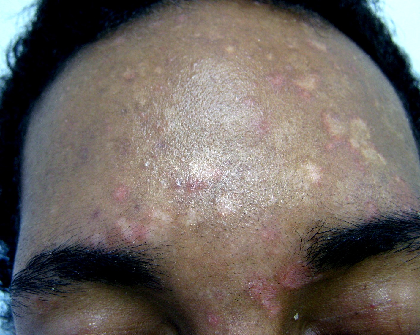
- Vitiligo: autoimmune (CD8⁺ T-cell) destruction of melanocytes → well-demarcated, chalk-white depigmented macules/patches, symmetric, favoring face/periorificial, hands, extensors; shows Koebner phenomenon. Biopsy: absent melanocytes. Linked to Hashimoto, T1DM, pernicious anemia, Addison, alopecia areata.
- Oculocutaneous albinism: normal melanocyte number, ↓melanin. OCA1 = tyrosinase deficiency; OCA2 = defective P-protein. Nystagmus, ↓visual acuity, photophobia; markedly ↑ risk of SCC/BCC (and melanoma, often amelanotic).
- Chédiak–Higashi: LYST mutation → giant lysosomal granules, partial (oculocutaneous) albinism, recurrent pyogenic infections, bleeding tendency, peripheral neuropathy.
- Pityriasis (tinea) versicolor: Malassezia → hypo- or hyperpigmented scaly macules on trunk that fail to tan (fungal azelaic acid inhibits tyrosinase). KOH: "spaghetti and meatballs."
- Ash-leaf macule: hypopigmented; earliest cutaneous sign of tuberous sclerosis (highlighted by Wood's lamp).
- Piebaldism: KIT mutation → congenital white forelock (poliosis) with stable depigmented patches.
Hypopigmentation comparison
| Condition | Mechanism | Melanocytes | Classic buzzword |
|---|---|---|---|
| Vitiligo | Autoimmune destruction | Absent | Chalk-white, Koebner, autoimmune associations |
| Albinism (OCA1) | Tyrosinase deficiency | Present (normal #) | Nystagmus, ↑↑ skin-cancer risk |
| Pityriasis versicolor | Malassezia; tyrosinase inhibition | Present | "Spaghetti & meatballs," doesn't tan |
| Ash-leaf macule | Developmental (TSC) | ↓ melanin | Tuberous sclerosis, Wood's lamp |
Vignette: A 28-year-old woman has enlarging, well-demarcated white patches on the dorsal hands and around the eyes and mouth over 6 months. The patches are completely depigmented, have no scale, and accentuate under Wood's lamp. She also reports fatigue and cold intolerance.
- Diagnosis: Vitiligo (autoimmune melanocyte destruction).
- Why: Depigmented (not scaly or merely hypopigmented) macules in a periorificial/acral distribution that Koebnerize.
- Next best step: Screen for associated autoimmune disease — TSH + anti-TPO (her symptoms suggest Hashimoto); also consider fasting glucose/HbA1c, B12, morning cortisol/ACTH given T1DM, pernicious anemia, and Addison associations.
- Management: Photoprotection, topical corticosteroids or calcineurin inhibitors, and narrowband UVB for extensive disease.

- Melasma ("chloasma"): symmetric brown patches on the face (cheeks, forehead, upper lip) driven by estrogen/progesterone (pregnancy, OCPs) plus UV. Treat: strict sun protection, hydroquinone, tretinoin.
- Café-au-lait macules: flat, uniform light-brown macules. ≥6 (>5 mm prepubertal, >15 mm postpubertal) suggests NF1, along with axillary/inguinal freckling (Crowe sign) and Lisch nodules.
- McCune–Albright: large, unilateral café-au-lait with jagged "coast of Maine" borders + polyostotic fibrous dysplasia + precocious puberty (mosaic GNAS activating mutation).
- Addison disease: ↓cortisol → ↑ACTH/POMC → MSH → diffuse hyperpigmentation of skin, mucosa, palmar creases, and scars.
- Acanthosis nigricans: velvety hyperpigmented flexural plaques; think insulin resistance or, if abrupt/extensive, visceral (gastric) adenocarcinoma.
Café-au-lait syndromes: NF1 vs. McCune–Albright
| Feature | NF1 | McCune–Albright |
|---|---|---|
| Gene | NF1 (neurofibromin, tumor suppressor) | GNAS (Gsα, mosaic activating) |
| CALM borders | Smooth — "coast of California" | Jagged — "coast of Maine" |
| Number/pattern | ≥6, scattered bilaterally | Often single, large, unilateral |
| Associations | Neurofibromas, Lisch nodules, freckling, optic glioma | Fibrous dysplasia, precocious puberty, endocrinopathies |
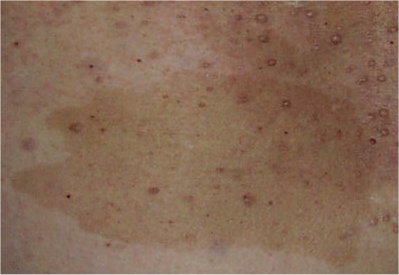
Vignette: A 19-year-old presents in summer with multiple oval, finely scaling macules on the upper trunk and shoulders — some lighter, some darker than surrounding skin. The lesions failed to tan with sun exposure and are mildly itchy.
- Most likely dx: Pityriasis (tinea) versicolor (Malassezia furfur/globosa).
- Next best step: KOH prep of the scale → "spaghetti and meatballs" (short hyphae + yeast clusters). Wood's lamp is optional and variable — when positive it shows yellow-gold to coppery-orange fluorescence (absent in many cases).
- Why the color change? Malassezia produces azelaic acid, which inhibits tyrosinase, blocking tanning in involved skin.
- Treatment: Topical selenium sulfide or ketoconazole; oral itraconazole/fluconazole for extensive disease. Pigment normalizes slowly over weeks to months (reassure — this is not scarring).
ABCDE — warning signs of melanoma (the pigmented lesion you must not miss):
- Asymmetry
- Border irregularity
- Color variegation
- Diameter >6 mm
- Evolution (change over time); some texts also cite Elevation
Next best step for a suspicious pigmented lesion: excisional biopsy with narrow margins (avoid shave biopsy — it can transect the lesion and preclude accurate staging) — Breslow depth is the single most important prognostic factor in localized disease. Risk factors: UV exposure, fair skin, dysplastic nevi. Superficial spreading = most common subtype; nodular = most aggressive; acral lentiginous = palms/soles/nails, the most common subtype in darker-skinned patients (and not UV-driven).
Practice Dermatology now
Board-style questions, spaced-repetition flashcards, and a Socratic AI tutor — free to start.