Pharyngeal Arch Derivatives & High-Yield Embryology
A boards-focused walkthrough of the pharyngeal apparatus — arches, pouches, clefts, and membranes — mapping each arch's cranial nerve, muscles, cartilage, and artery to the classic Step 1 syndromes (DiGeorge, Treacher Collins, branchial cleft cyst) and the recurrent laryngeal nerve.
The pharyngeal apparatus at a glance
The pharyngeal (branchial) apparatus builds the face, neck, and many head and foregut derivatives. It is made of 6 pharyngeal arches (the 5th is rudimentary/absent in humans, so boards count 1, 2, 3, 4, 6), pharyngeal pouches (endoderm-lined, internal), pharyngeal clefts/grooves (ectoderm-lined, external), and pharyngeal membranes (where ectoderm meets endoderm).
Each arch is a self-contained unit built from all three germ layers plus neural crest, and each keeps its own cartilage, muscle, cranial nerve, and aortic arch artery — even if the muscle later migrates far from the arch. Learn the arch → nerve → muscle → cartilage map and you can predict nearly every associated syndrome tested on Step 1.
- Cranial nerve by arch: 1 = CN V (V2/V3), 2 = CN VII, 3 = CN IX, 4 = CN X (superior laryngeal), 6 = CN X (recurrent laryngeal).
- Arch 1 → muscles of mastication (+ mylohyoid, anterior digastric, tensor tympani, tensor veli palatini); Meckel cartilage → Malleus, Incus, Mandible.
- Arch 2 → muscles of facial expression (+ stapedius, stylohyoid, posterior digastric, platysma); Reichert cartilage → Stapes, Styloid process, lesser horn + upper body of hyoid, stylohyoid ligament.
- Arch 3 → stylopharyngeus (the only CN IX muscle); cartilage → greater horn + lower body of hyoid.
- Arch 4 → most pharyngeal constrictors, cricothyroid, levator veli palatini; Arch 6 → all intrinsic laryngeal muscles EXCEPT cricothyroid. Arches 4 & 6 form the laryngeal cartilages (thyroid, cricoid, arytenoid, corniculate, cuneiform).
- Germ-layer origins (tested): arch cartilage, bone & connective tissue = neural crest; arch muscles = mesoderm (myoblasts from somitomeres/paraxial mesoderm). The arch's cranial nerve migrates with its muscle wherever it goes — so the nerve tells you the source arch.
Pharyngeal arch derivatives
| Arch | Cranial nerve | Muscles | Cartilage / skeletal | Artery |
|---|---|---|---|---|
| 1 (mandibular) | CN V2 / V3 | Mastication (temporalis, masseter, pterygoids), mylohyoid, ant. digastric, tensor tympani, tensor veli palatini | Malleus, incus, mandible, sphenomandibular ligament (Meckel) | Part of maxillary artery |
| 2 (hyoid) | CN VII | Facial expression, stapedius, stylohyoid, post. digastric, platysma | Stapes, styloid process, lesser horn + upper hyoid, stylohyoid ligament (Reichert) | Stapedial + hyoid arteries |
| 3 | CN IX | Stylopharyngeus | Greater horn + lower body of hyoid | Common carotid; proximal internal carotid |
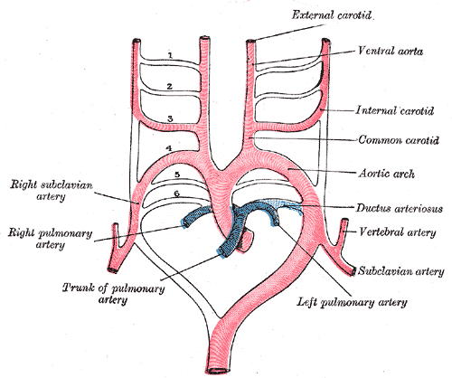
| 4 | CN X (superior laryngeal) | Most pharyngeal constrictors, cricothyroid, levator veli palatini | Thyroid, cricoid, arytenoid, corniculate, cuneiform | L → aortic arch; R → proximal right subclavian |
| 6 | CN X (recurrent laryngeal) | All intrinsic laryngeal muscles except cricothyroid | (shares laryngeal cartilages) | Proximal pulmonary arteries; L → ductus arteriosus |

Arch cranial nerves (1 → 2 → 3 → 4 → 6): CN V, VII, IX, X — the branchiomotor (pharyngeal-arch) cranial nerves, in numeric order (arches 4 and 6 both run on CN X).
1st arch = the "M" arch: Malleus, incus, Mandible, Meckel cartilage, Muscles of Mastication, Mylohyoid.
2nd arch = the "S" arch (Reichert): Stapes, Styloid process, Stylohyoid ligament, leSser horn of hyoid; muscles Stapedius, Stylohyoid.
Laryngeal nerves: arch 4 → superior laryngeal (cricothyroid), arch 6 → recurrent laryngeal (everything else). "Cricothyroid is the odd one out."
Pouches, clefts, and membranes
Where arches give skeletal and muscular derivatives, the endoderm-lined pouches (1–4) form glandular and epithelial structures.
A classic boards trap lives here: the inferior parathyroids arise from the 3rd pouch — they migrate farther caudally, dragged down by the descending thymus — while the superior parathyroids arise from the 4th pouch. The higher pouch ends up in the lower position.
The ectoderm-lined clefts (grooves) are external. Only the 1st cleft persists (→ external auditory meatus); clefts 2–4 are normally obliterated within the cervical sinus, and failure to obliterate leaves a branchial cleft cyst. The pharyngeal membranes contribute the tympanic membrane (1st membrane), the meeting point of pouch 1, cleft 1, and their arches.
- Pouch 1 → middle ear cavity, auditory (Eustachian) tube, mastoid air cells (+ tympanic membrane at the 1st membrane).
- Pouch 2 → epithelial lining of the palatine tonsil crypts.
- Pouch 3 (dorsal) → inferior parathyroids; Pouch 3 (ventral) → thymus.
- Pouch 4 (dorsal) → superior parathyroids; Pouch 4 (ventral) → ultimobranchial body → parafollicular (C) cells of thyroid (calcitonin; the C cells themselves seed in from neural crest).
- Clefts: 1st → external auditory meatus; 2nd–4th → obliterated. Persistence → branchial cleft cyst (lateral neck, anterior to SCM).
- Parathyroid trap: 3rd pouch → inferior, 4th pouch → superior ("3 ends up below 4").

- DiGeorge syndrome (22q11.2 deletion): failed 3rd + 4th pouch development → thymic aplasia (T-cell deficiency, recurrent viral/fungal infection) + parathyroid aplasia (hypocalcemia, tetany, seizures) + conotruncal cardiac defects (tetralogy of Fallot, truncus arteriosus — neural crest fails to septate the outflow tract) + abnormal facies/cleft palate. Mnemonic: CATCH-22.
- Treacher Collins syndrome: 1st arch neural crest fails to migrate → mandibular + zygomatic hypoplasia, downslanting palpebral fissures, ear anomalies with conductive hearing loss.
- Branchial (2nd) cleft cyst: painless, fluctuant lateral neck mass anterior to the sternocleidomastoid; persistent cervical sinus.
- Recurrent laryngeal nerve (arch 6): on the right it loops under the right subclavian artery (arch 4); on the left it loops under the aortic arch / ligamentum arteriosum (arch 6) — so the left RLN descends into the thorax and is vulnerable to a dilated left atrium (mitral stenosis), aortic aneurysm, or lung apex tumor → hoarseness (Ortner / cardiovocal syndrome).
- MEN 2A/2B: medullary thyroid carcinoma from parafollicular C cells (4th pouch) → elevated calcitonin.
Neck cyst mimics — branchial cleft vs thyroglossal duct
| Feature | Branchial cleft cyst | Thyroglossal duct cyst |
|---|---|---|
| Origin | Persistent 2nd cleft / cervical sinus | Persistent thyroglossal duct (thyroid descent from foramen cecum) |
| Location | Lateral neck, anterior to SCM | Midline neck |
| Moves with tongue protrusion / swallowing | No | Yes (elevates) |
| Lining / contents | Squamous epithelium + lymphoid tissue | May contain ectopic thyroid tissue |
| Key association | Painless lateral mass in young adult | Foramen cecum; may be the only functioning thyroid tissue |
- Anterior 2/3 mucosa = 1st arch: general sensation CN V3, taste CN VII (chorda tympani).
- Posterior 1/3 = 3rd (+ 4th) arch: general sensation and taste CN IX; extreme posterior/epiglottic region CN X.
- Motor: all tongue muscles = CN XII (from occipital myotomes) — EXCEPT palatoglossus = CN X.
- Thyroid descends from the foramen cecum; failed descent → lingual thyroid or thyroglossal duct cyst.
Practice Embryology now
Board-style questions, spaced-repetition flashcards, and a Socratic AI tutor — free to start.