Peripheral Neuropathy & Guillain-Barré
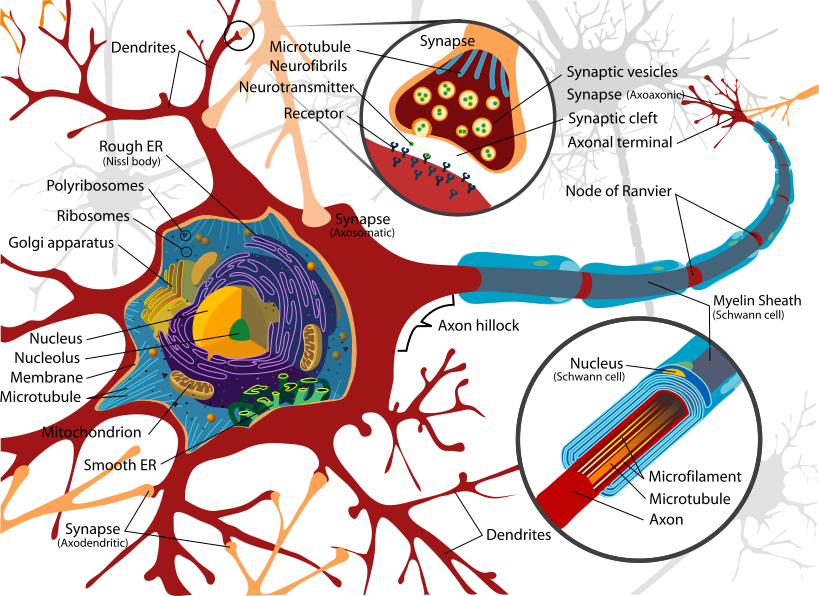
A high-yield boards lesson on peripheral neuropathy patterns and Guillain-Barré syndrome, moving from pathophysiology and localization through CSF/NCS diagnosis to the emergency management decisions (respiratory monitoring, IVIG vs plasma exchange, and the classic steroid trap). Includes a GBS-vs-CIDP comparison, Miller Fisher recognition, and real vignette buzzwords with next-best-step logic. Reviewed and corrected for clinical accuracy: the myelin image was changed from a CNS oligodendrocyte to a Schwann-cell (peripheral) diagram to match GBS localization.
Pathophysiology & Classification
Peripheral neuropathy is dysfunction of peripheral nerves, classified two ways that drive the differential: by pattern and by primary pathology (axonal vs demyelinating).
- Distal symmetric polyneuropathy ("stocking-glove," length-dependent): usually metabolic/toxic — diabetes (#1 in the developed world), alcohol, B12 deficiency, uremia, chemotherapy.
- Mononeuropathy: focal compression/entrapment (median → carpal tunnel; common peroneal at the fibular head → foot drop).
- Mononeuritis multiplex: several named nerves affected stepwise — think vasculitis and diabetes; leprosy worldwide.
Guillain-Barré syndrome (GBS) is the prototypical acute immune polyradiculoneuropathy. Days to weeks after infection, molecular mimicry generates autoantibodies against nerve gangliosides, producing demyelination (AIDP, the most common Western subtype) or axonal damage (AMAN, anti-GD1a/GM1). GBS is a neurologic emergency: respiratory and autonomic failure are the killers, and immunotherapy is time-sensitive.
- Diabetes = most common peripheral neuropathy (developed world); leprosy = classic answer for most common worldwide
- B12 deficiency → distal sensory loss plus dorsal-column/corticospinal signs (subacute combined degeneration): neuropathy WITH upper-motor-neuron signs; ↑ methylmalonic acid & homocysteine
- NCS pattern: axonal = reduced amplitude; demyelinating = slowed conduction velocity, conduction block, prolonged/absent F-waves
- GBS = ascending symmetric weakness + areflexia, often days after infection — Campylobacter jejuni (#1 overall), CMV (most common viral), EBV, Mycoplasma, Zika; also post-surgery/vaccination
- CSF: albuminocytologic dissociation (↑ protein, normal cell count); may be normal in week 1. Pleocytosis (>50 WBCs) → look for HIV, Lyme, or malignancy
- Treatment = IVIG or plasma exchange (equally effective; do not combine); corticosteroids do NOT work in GBS (they DO help CIDP)
- Miller Fisher variant: ophthalmoplegia + ataxia + areflexia, anti-GQ1b antibodies
Vignette: A 32-year-old man develops tingling in the feet, then progressive, symmetric leg weakness ascending to the trunk and arms over 4 days. Two weeks earlier he had bloody diarrhea. Exam: symmetric flaccid weakness, absent deep tendon reflexes, minimal sensory loss.
Diagnosis: Guillain-Barré syndrome (AIDP), post-Campylobacter.
Next best step (do NOT jump straight to the LP):
- Assess airway/breathing — bedside FVC and negative inspiratory force (NIF), trended serially. Respiratory failure is the leading cause of death.
- Continuous cardiac telemetry for dysautonomia (arrhythmias, labile BP).
- Confirm with LP (albuminocytologic dissociation) and nerve conduction studies (earliest finding: prolonged/absent F-waves).
- Start IVIG or plasma exchange.
Buzzwords: ascending paralysis, areflexia, recent diarrheal illness, elevated CSF protein with normal cell count.

GBS (AIDP) vs CIDP
| Feature | GBS / AIDP | CIDP |
|---|---|---|
| Tempo to nadir | Acute, ≤4 weeks | Chronic, >8 weeks (progressive/relapsing) |
| Trigger | Post-infectious (Campylobacter) | Usually none |
| Reflexes | Areflexia | Hypo-/areflexia |
| CSF | Albuminocytologic dissociation | Albuminocytologic dissociation |
| NCS | Demyelinating | Demyelinating |
| Corticosteroids | Ineffective | First-line, effective |
| First-line Rx | IVIG or plasma exchange | Steroids, IVIG, or plasma exchange |
Diagnosis is clinical, supported by:
- LP: albuminocytologic dissociation. Often normal early — a normal week-1 tap does not exclude GBS.
- Nerve conduction studies: most useful early finding is prolonged or absent F-waves / H-reflexes (proximal nerve-root demyelination); conduction block and slowing appear later.
- MRI spine with contrast: may show enhancement of the anterior (ventral) nerve roots / cauda equina; mainly used to exclude cord compression or transverse myelitis mimics.
Red flags against GBS (hunt for another cause):
- Sensory level, early bowel/bladder involvement, or UMN signs → cord compression / transverse myelitis.
- Descending weakness with diplopia, dry mouth, dilated pupils → botulism.
- Also consider tick paralysis, heavy-metal toxicity, hypokalemia, and acute intermittent porphyria.
Treat GBS as a neuro-ICU-level emergency.
- Respiratory monitoring — the 20/30/40 rule → consider elective intubation when FVC < 20 mL/kg, maximal inspiratory pressure weaker than −30 cm H2O, or maximal expiratory pressure < 40 cm H2O. Intubate early — do not wait for hypoxia/hypercapnia (late and dangerous).
- Dysautonomia: continuous telemetry; treat arrhythmias; use vasoactive drugs cautiously (exaggerated, labile responses).
- Immunotherapy: IVIG or plasma exchange, ideally within ~2 weeks of onset — equally effective, never combined. No corticosteroids.
- Supportive: VTE prophylaxis (immobility), neuropathic pain control (gabapentin), physical therapy, and watch for SIADH.
Most patients recover over weeks-to-months; mortality (~3-7%) is driven by respiratory and autonomic complications.
Causes of polyneuropathy — "DANG THERAPIST":
- D — Diabetes
- A — Alcohol
- N — Nutritional (B12, thiamine, B6)
- G — Guillain-Barré
- T — Toxins/drugs (vincristine, cisplatin, isoniazid, heavy metals)
- H — Hereditary (Charcot-Marie-Tooth)
- E — Endocrine (hypothyroidism)
- R — Rheumatologic/vasculitis (RA, SLE, PAN)
- A — Amyloidosis
- P — Paraneoplastic / Porphyria
- I — Infectious (HIV, Lyme, leprosy, diphtheria)
- S — Sarcoidosis
- T — Tumor / compression
Miller Fisher triad — the 3 core signs: Ophthalmoplegia + Ataxia + Areflexia (anti-GQ1b).
Vignette: A 40-year-old woman, 10 days after an upper-respiratory illness, reports double vision, an unsteady gait, and clumsy fine movements. Exam: ophthalmoplegia, limb and gait ataxia, and absent reflexes — with normal strength and near-normal sensation.
Diagnosis: Miller Fisher syndrome, the GBS variant defined by ophthalmoplegia + ataxia + areflexia.
Next best step / pearls:
- Send anti-GQ1b antibodies (positive in ~85-90%).
- Still monitor for respiratory/autonomic involvement and overlap with classic GBS.
- CSF again shows albuminocytologic dissociation.
- Treat like GBS (IVIG or plasma exchange) if progressive; many cases are self-limited.
Distinguish from: brainstem stroke (long-tract signs), botulism (descending, pupil-involving), and Wernicke (ophthalmoplegia + ataxia + confusion, reflexes intact).
Practice Neuro now
Board-style questions, spaced-repetition flashcards, and a Socratic AI tutor — free to start.