Pericarditis, Effusion & Cardiac Tamponade
A boards-focused walkthrough of the pericardial disease spectrum — acute pericarditis, effusion, tamponade, and constriction — emphasizing classic vignette buzzwords, ECG/echo findings, and next-best-step decisions. Includes three high-yield vignettes and a side-by-side comparison table.
The pericardial disease continuum
The pericardium is a two-layer sac (visceral + parietal) holding ~15–50 mL of serous fluid. Inflammation — acute pericarditis — irritates these layers, producing pleuritic pain and a friction rub. Injury or inflammation can exude fluid into the sac (pericardial effusion). What matters hemodynamically is not the absolute volume but the rate of accumulation: the stiff parietal pericardium slowly stretches to accommodate 1–2 L, but rapid collection of as little as ~150 mL sharply raises intrapericardial pressure. Once that pressure exceeds cardiac filling pressures, diastolic filling is impaired, stroke volume falls, and cardiac tamponade — a form of obstructive shock — develops. Over time, a scarred, non-compliant pericardium can restrict filling as constrictive pericarditis. Boards test this continuum end to end: recognize the pain/rub/ECG of pericarditis, treat tamponade as an emergency, and separate constriction from restrictive cardiomyopathy.
- Diagnosis = ≥2 of 4: pleuritic chest pain, pericardial friction rub, characteristic ECG changes, new/worsening pericardial effusion
- Pain: sharp, pleuritic, worse when supine and on inspiration, relieved by sitting up and leaning forward
- Friction rub: scratchy, often triphasic; loudest at the left lower sternal border, leaning forward, end-expiration
- ECG: diffuse, concave-up ST elevation with PR depression; PR elevation (with ST depression) in aVR — separates it from STEMI (regional, convex, with reciprocal changes)
- Most common cause: viral/idiopathic (classically Coxsackievirus B). Others: uremia, post-MI (Dressler), autoimmune (SLE, RA), TB, malignancy, radiation
- Labs: ↑ESR/CRP; a rising troponin suggests myopericarditis
- Treatment: NSAID (ibuprofen or high-dose aspirin) + colchicine; colchicine reduces recurrence. Avoid steroids first-line (↑recurrence)
- Post-MI pericarditis: prefer aspirin, avoid other NSAIDs/steroids (impair infarct healing). Uremic pericarditis: treat with dialysis, not NSAIDs
Vignette: A 28-year-old man has 3 days of sharp substernal chest pain that worsens when he lies flat and eases when he leans forward. He had a URI last week. A scratchy sound is heard at the left sternal border. ECG shows diffuse ST-segment elevation and PR-segment depression; troponin is normal.
Diagnosis: Acute (viral/idiopathic) pericarditis.
Next best step: Start NSAID (ibuprofen or aspirin) + colchicine and restrict strenuous activity. Obtain echocardiography to look for an effusion.
Trap: Diffuse, concave-up ST elevation with PR depression — do not reflexively activate the cath lab. STEMI ST elevation is regional and convex with reciprocal depression. If the troponin were elevated, call it myopericarditis (still get an echo; involve cardiology).

- Effusion ECG: low voltage and electrical alternans (beat-to-beat swing in QRS amplitude = heart swinging in fluid)
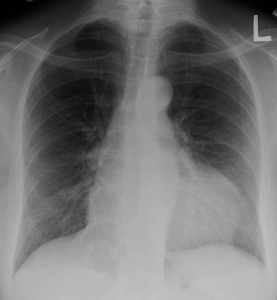
- CXR: 'water-bottle' cardiac silhouette (needs ≳200 mL); lung fields clear
- Echocardiography = test of choice for both effusion and tamponade
- Tamponade physiology: ↑intrapericardial pressure → equalization of diastolic pressures, impaired filling, ↓cardiac output → obstructive shock
- Beck's triad: hypotension + JVD + muffled/distant heart sounds
- Pulsus paradoxus: inspiratory fall in systolic BP >10 mmHg (also in severe asthma/COPD and tension pneumothorax)
- JVP: prominent x descent with blunted/absent y descent (contrast with constriction: prominent x and y)
- Echo signs: RA collapse (systolic) and RV free-wall collapse (early diastolic), dilated IVC (plethora), exaggerated respiratory variation of transvalvular inflow
- Kussmaul sign (JVP rises with inspiration) points to constriction, not classic tamponade
Vignette: A 60-year-old woman with metastatic breast cancer is dyspneic and cool. BP 82/60, HR 120, JVP elevated, heart sounds distant. Systolic BP drops 18 mmHg on inspiration. ECG shows sinus tachycardia, low voltage, and electrical alternans.
Diagnosis: Cardiac tamponade (malignant effusion).
Next best step:
- Urgent bedside echocardiography to confirm (RV diastolic collapse, IVC plethora).
- Cautious IV fluids to support preload while arranging drainage.
- Echo-guided pericardiocentesis — definitive. For malignant/recurrent effusions → pericardial window.
Avoid: diuretics, vasodilators, and positive-pressure ventilation — all drop preload and can precipitate arrest.
Key exception: If tamponade is from a type A aortic dissection or ventricular free-wall rupture, the answer is emergency surgery — pericardiocentesis can trigger fatal re-bleeding and is not definitive.
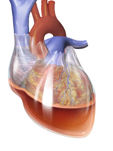
Beck's triad of acute tamponade — the 3 D's:
- Distant (muffled) heart sounds
- Distended neck veins (JVD)
- Decreased arterial pressure (hypotension)
Pulsus paradoxus = exaggerated (>10 mmHg) inspiratory drop in systolic BP. Remember the big three causes: tamponade, severe asthma/COPD, and tension pneumothorax.
Pericarditis ECG — 'PR down, ST up, diffusely': PR depression + concave-up ST elevation across most leads (with PR elevation in aVR).
Acute pericarditis vs tamponade vs constriction
| Feature | Acute pericarditis | Cardiac tamponade | Constrictive pericarditis |
|---|---|---|---|
| Onset | Acute | Acute / subacute | Chronic |
| Hallmark exam | Friction rub | Beck's triad, pulsus paradoxus | Kussmaul sign, pericardial knock |
| JVP waveform | — | Prominent x, absent y | Prominent x and y (rapid y) |
| ECG | Diffuse ST↑, PR↓ | Low voltage, electrical alternans | Low voltage, ± atrial fibrillation |
| Imaging | Echo: ± effusion | Echo: RA (systolic) + RV (diastolic) collapse, IVC plethora | CT/CXR: thick/calcified pericardium; cath 'square-root' sign |
| Treatment | NSAID + colchicine | Pericardiocentesis (± window) | Pericardiectomy |
Vignette: A 55-year-old man, 2 years after treated TB pericarditis (or prior cardiac surgery/radiation), has progressive dyspnea, JVD that rises with inspiration (Kussmaul sign), hepatomegaly, ascites, and leg edema. An early diastolic pericardial knock is heard. CT shows a thickened, calcified pericardium.
Diagnosis: Constrictive pericarditis.
Workup: Echo and cardiac catheterization reveal equalization of diastolic pressures and a 'dip-and-plateau' (square-root) sign.
Definitive treatment: Pericardiectomy (diuretics only palliate congestion).
Key distinction — constriction vs restrictive cardiomyopathy: pericardial calcification/thickening and respiratory discordance of LV/RV pressures (ventricular interdependence) favor constriction (surgically curable); marked biatrial enlargement, high BNP, and intrinsic myocardial disease favor restriction.
Practice Cardiology now
Board-style questions, spaced-repetition flashcards, and a Socratic AI tutor — free to start.