Pelvis & Perineum Anatomy
A board-focused tour of pelvis and perineum anatomy: the pelvic floor and perineal pouches, the all-important pectinate line, the pudendal nerve and its blocks, urethral rupture patterns, and autonomic control of continence and sexual function — anchored to the clinical vignettes Step 1 loves.
The Big Picture
The pelvis is the funnel-shaped space bounded by the bony pelvis and closed inferiorly by the pelvic diaphragm (levator ani + coccygeus). Below the pelvic diaphragm lies the perineum, a diamond split by an imaginary line between the two ischial tuberosities into an anterior urogenital triangle (external genitalia, urethra, and — in the male — the root of the penis) and a posterior anal triangle (anal canal + ischioanal fossae).
Boards rarely ask you to name every ligament — they test relationships and consequences: the pectinate line and its portal–caval watershed, the pudendal nerve and where it is blocked, urethral rupture patterns, autonomic control of erection/ejaculation, and the ureter's vulnerable course. Orient yourself to three supply lines: blood below the pelvic brim comes mainly from the internal iliac artery; somatic innervation of the perineum is the pudendal nerve (S2–S4); and autonomics arrive via sympathetic hypogastric nerves (T10–L2) and parasympathetic pelvic splanchnics (S2–S4).
- Pelvic diaphragm = levator ani (puborectalis + pubococcygeus + iliococcygeus) + coccygeus. It supports the viscera and maintains continence; the puborectalis sling creates the anorectal angle and relaxes to permit defecation.
- Perineal body = fibromuscular central tendon just anterior to the anus; it anchors the external anal sphincter, bulbospongiosus, and the transverse perineal muscles. Torn in childbirth → pelvic organ prolapse/incontinence (the rationale for episiotomy).
- Two urogenital pouches: the deep perineal pouch holds the external urethral sphincter and membranous urethra (+ bulbourethral/Cowper glands in males, whose ducts drain into the spongy urethra below); the superficial pouch holds the erectile bodies, their muscles (ischiocavernosus, bulbospongiosus), and the greater vestibular (Bartholin) glands in females.
- Internal iliac artery supplies the pelvis; its internal pudendal branch is the artery of the perineum (→ inferior rectal, perineal, and dorsal artery of the penis/clitoris).
- Autonomics: sympathetic = hypogastric nerves / superior hypogastric plexus (T10–L2); parasympathetic = pelvic splanchnic nerves (S2–S4).
- Gonadal veins: the right ovarian/testicular vein drains straight into the IVC; the left drains into the left renal vein → left-sided varicocele (a new, non-decompressing left varicocele in an older man = renal cell carcinoma invading the renal vein until proven otherwise).

The Pectinate (Dentate) Line — the single most tested comparison
| Feature | Above pectinate line | Below pectinate line |
|---|---|---|
| Embryonic origin | Endoderm (hindgut) | Ectoderm (proctodeum) |
| Epithelium | Simple columnar | Stratified squamous |
| Arterial supply | Superior rectal a. (from IMA) | Inferior rectal a. (from internal pudendal) |
| Venous drainage | Superior rectal → IMV → portal vein | Inferior rectal → internal pudendal → internal iliac → IVC |
| Innervation | Visceral (hypogastric plexus) — insensate to pain | Somatic (inferior rectal n.) — pain-sensitive |
| Lymphatics | Internal iliac nodes | Superficial inguinal nodes |
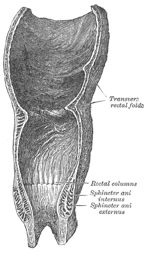
| Hemorrhoids | Internal — painless | External — painful |
Straddle injury (a man slips and lands astride a bike crossbar or fence): ruptures the bulbous (spongy) urethra in the superficial perineal pouch. Urine and blood escape into the superficial perineal space and — because Colles fascia is continuous with the dartos and abdominal Scarpa fascia — track into the scrotum, penis, and up the anterior abdominal wall, but not into the thighs (Colles fascia fuses to the ischiopubic rami and fascia lata). Clue: blood at the meatus with scrotal/penile swelling and bruising.
Pelvic fracture (high-energy trauma, e.g., MVC): shears the membranous urethra — the least distensible segment, passing through the deep perineal pouch — so urine collects in the deep perineal space / extraperitoneal (retropubic) space of Retzius. Clues: blood at the meatus, a high-riding, boggy prostate, and inability to void.
Order a retrograde urethrogram before catheterizing any suspected urethral injury.
The pudendal nerve (S2–S4) leaves the pelvis through the greater sciatic foramen, hooks around the ischial spine / sacrospinous ligament, and re-enters through the lesser sciatic foramen into the pudendal (Alcock) canal on the lateral wall of the ischioanal fossa. Its branches in order: inferior rectal → perineal → dorsal nerve of the penis/clitoris.
Vignette — pudendal block: during labor, a needle is guided to the ischial spine (palpated transvaginally) to anesthetize the perineum for delivery or episiotomy. Chronic entrapment (cyclists, prolonged sitting) → perineal pain, numbness, and sexual dysfunction; bilateral injury → fecal/urinary incontinence because the external anal and urethral sphincters lose their motor supply.
Vignette — ischioanal fossa: this fat-filled, wedge-shaped space flanking the anal canal is a classic site for an anorectal (ischioanal) abscess; infection can burrow posteriorly behind the anal canal into the opposite fossa to form a horseshoe abscess.

Pelvic & Perineal Nerves — root, function, lesion
| Nerve | Roots | Function | Lesion / deficit |
|---|---|---|---|
| Pudendal | S2–S4 | Motor: external anal & urethral sphincters, perineal muscles. Sensory: perineum & external genitalia | Fecal/urinary incontinence, perineal anesthesia, sexual dysfunction; blocked at the ischial spine |
| Pelvic splanchnic | S2–S4 (parasympathetic) | Erection; detrusor contraction (voiding); motility distal to the splenic flexure | Erectile dysfunction, neurogenic (atonic) bladder |
| Sympathetic (hypogastric) | T10–L2 | Emission + internal urethral sphincter closure; ejaculation coordination | Retrograde ejaculation, failure of emission |
| Nerve to levator ani | S3–S4 | Motor to the pelvic diaphragm | Pelvic floor weakness → prolapse, stress incontinence |
| Obturator | L2–L4 | Motor: thigh adductors. Sensory: medial thigh | Weak adduction + medial thigh numbness; injured in pelvic surgery, difficult labor, or obturator hernia |
- "S2, S3, S4 keep the pee (and poop) off the floor." The pudendal nerve roots and the pelvic floor: continence at the external urethral and anal sphincters depends on S2–S4.
- "Point and Shoot." Parasympathetic (pelvic splanchnics) = Point → erection (NO-mediated helicine artery dilation, the PDE5-inhibitor target). Sympathetic = Shoot → emission (semen into the prostatic urethra) + closure of the internal urethral sphincter to block retrograde flow; the final expulsion is a somatic reflex (pudendal-driven rhythmic bulbospongiosus contraction). α-blockers or sympathetic injury → retrograde ejaculation.
- "Water under the bridge." The ureter (water) passes inferior to the uterine artery (bridge) ~2 cm lateral to the cervix (in the male, the ductus deferens crosses over the ureter). This is why the ureter is easily clamped or ligated during hysterectomy.
- Suspensory (infundibulopelvic) ligament carries the ovarian vessels to/from the aorta and IVC; it is ligated during oophorectomy — and the ureter runs just beneath it at the pelvic brim, at risk of injury.
- Cardinal (transverse cervical) ligament = the chief ligamentous support of the uterus; it carries the uterine artery (with the ureter passing beneath).
- Round ligament of the uterus = gubernaculum remnant; it travels through the inguinal canal to the labia majora (so this region drains to superficial inguinal nodes) and causes round-ligament pain in pregnancy — it provides little true support.
- Rectouterine pouch of Douglas = the most dependent recess of the female peritoneal cavity, so blood or pus pools here (ruptured ectopic, PID) and can be sampled by culdocentesis. The male equivalent is the rectovesical pouch.
Practice Anatomy now
Board-style questions, spaced-repetition flashcards, and a Socratic AI tutor — free to start.