Pelvic Pain & Adnexal Masses
A Step 2 CK high-yield walkthrough of acute pelvic pain and adnexal masses, built as a decision tree (β-hCG first → acute vs chronic → TVUS → stability → age/malignancy risk) with next-best-step vignettes for ovarian torsion and ectopic pregnancy.
The Pelvic Pain Decision Tree
Pelvic pain and adnexal masses are tested as a decision tree, and the first branch is always the same: check a urine or serum β-hCG. Pregnancy reframes the entire differential (ectopic, corpus luteum) and gates later choices like methotrexate. The second question is acute vs chronic: sudden, severe, unilateral pain points to torsion, ectopic, or ruptured cyst; cyclic or chronic pain suggests endometriosis, dysmenorrhea, or a slowly enlarging mass. Transvaginal ultrasound (TVUS) is the imaging workhorse for both — it characterizes size, cystic vs solid, septations, and Doppler flow. Hemodynamic stability sets the tempo: an unstable patient with a positive β-hCG and free fluid goes to the OR, not the CT scanner. Finally, age drives malignancy risk — functional cysts dominate the reproductive years, but a complex or solid adnexal mass in a premenarchal or postmenopausal patient is cancer until proven otherwise.
- β-hCG first in any reproductive-age woman with pelvic pain — no exceptions.
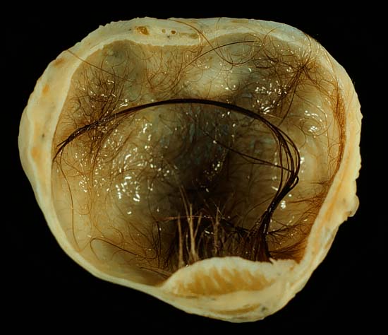
- Ovarian torsion = sudden severe unilateral pain + nausea/vomiting, usually with a mass/cyst >5 cm (mature teratoma is the classic lead point).
- Doppler may show reduced flow, but normal flow does NOT exclude torsion (ovary has a dual blood supply). It is a clinical/surgical diagnosis.
- Torsion → laparoscopy with detorsion + ovarian preservation; don't delay for repeat imaging or default to oophorectomy in a young patient.
- Ectopic: empty uterus on TVUS when β-hCG is above the discriminatory zone (~1,500–2,000); a normal IUP roughly doubles β-hCG every 48 h — a plateaued/subnormal rise flags ectopic or nonviable.
- Ruptured cyst (hemorrhagic corpus luteum): sudden pain post-coitus/exercise + free fluid → manage conservatively if stable.
- Tubo-ovarian abscess: PID + fever + adnexal mass → IV antibiotics; drain if large or not improving.
- Malignancy red flags: solid components, thick septations, ascites, bilaterality, ↑CA-125, postmenopausal → refer to gyn-onc; do not aspirate.
Acute Pelvic Pain: Rapid Differential
| Diagnosis | Classic buzzwords | Best test | Next best step |
|---|---|---|---|
| Ovarian torsion | Sudden severe unilateral pain, N/V, mass/cyst >5 cm | TVUS + Doppler (whirlpool sign, ↓flow) | Emergent laparoscopy → detorse + preserve ovary |
| Ectopic pregnancy | Amenorrhea, pain, spotting; +β-hCG, empty uterus | TVUS + serial β-hCG | MTX if stable/unruptured; salpingectomy if unstable |
| Ruptured ovarian cyst | Sudden pain after sex/exercise, free fluid | TVUS | Conservative if stable; surgery if unstable |
| Tubo-ovarian abscess | Fever, discharge, CMT, adnexal mass, recent PID | TVUS/CT, +NAAT | IV antibiotics; drain if >7 cm or no response |
| Appendicitis | Migratory periumbilical → RLQ pain, anorexia, low-grade fever; β-hCG negative | CT abdomen/pelvis (US/MRI if pregnant) | Appendectomy |
Vignette: A 24-year-old woman has sudden, severe right-lower-quadrant pain that woke her from sleep, with nausea and two episodes of vomiting. She is afebrile and β-hCG is negative. TVUS shows a 6 cm right adnexal mass with an enlarged, edematous ovary and diminished venous Doppler flow.
Diagnosis: Ovarian torsion — a mature cystic teratoma is the classic lead point.
Next best step: Emergent diagnostic laparoscopy with detorsion and ovarian conservation (cystectomy of the underlying mass). Do not wait for repeat imaging, and do not default to oophorectomy in a young patient.
Board trap: Preserved arterial/venous flow on Doppler does NOT rule out torsion, because the ovary receives blood from both the ovarian artery and the ovarian branch of the uterine artery. If suspicion is high, take her to the OR.
Vignette: A 28-year-old woman with prior chlamydial PID and 7 weeks of amenorrhea has unilateral pelvic pain and spotting. She is hemodynamically stable. β-hCG is 1,800; TVUS shows an empty uterus with a 3 cm right tubal mass, no free fluid, and no fetal cardiac activity.
Diagnosis: Unruptured tubal ectopic — β-hCG above the discriminatory zone with no intrauterine gestation.
Next best step: Single-dose IM methotrexate. She qualifies: stable, unruptured, β-hCG <5,000, small mass, no fetal cardiac activity, and able to follow up. Check β-hCG on day 4 and day 7 and expect a ≥15% decline between them; if not, give a second dose.
Switch to surgery (salpingectomy, or salpingostomy to preserve the tube) if she is unstable, ruptured, or has an MTX contraindication (hepatic/renal disease, immunodeficiency, breastfeeding, active pulmonary/peptic ulcer disease).
- Reproductive age = mostly benign: functional cysts (follicular, corpus luteum), endometrioma ("chocolate cyst," homogeneous ground-glass echoes), mature cystic teratoma/dermoid (most common benign neoplasm; classic torsion lead point).
- Simple cyst <5 cm, premenopausal: reassure; no follow-up needed (functional cyst → confirm resolution with repeat US in ~6–8 weeks).
- Premenarchal or postmenopausal complex/solid mass: malignancy until proven otherwise.
- CA-125 is most useful in postmenopausal women — premenopausally it is falsely raised by endometriosis, fibroids, PID, menstruation, and pregnancy.
- Refer to gyn-onc for solid/complex mass, ascites, ↑CA-125, or a high risk-of-malignancy index — do not aspirate (risk of spill/upstaging).
- Epithelial tumors are the most common ovarian cancers overall; germ cell tumors predominate in patients <20.
Board buzzwords worth memorizing cold:
- Dysgerminoma → LDH — most common malignant germ cell tumor; female counterpart of seminoma.
- Yolk sac (endodermal sinus) → AFP — Schiller–Duval bodies; young girls.
- Choriocarcinoma → β-hCG (very high) — theca-lutein cysts.
- Granulosa cell → inhibin + estrogen → precocious puberty or postmenopausal bleeding (Call–Exner bodies).
- Sertoli–Leydig → testosterone → virilization (Reinke crystals).
- Fibroma → Meigs syndrome = ovarian fibroma + ascites + pleural effusion (resolves after tumor removal).
- Serous cystadenocarcinoma → psammoma bodies; CA-125 tracks epithelial cancers.
Practice OB/GYN now
Board-style questions, spaced-repetition flashcards, and a Socratic AI tutor — free to start.