Pediatric Eye Disease (Strabismus, Retinoblastoma, ROP)
A high-yield board review of the three exam-favorite pediatric eye diseases — strabismus/amblyopia, retinoblastoma, and retinopathy of prematurity — anchored on the two red-flag signs (leukocoria and strabismus) with vignette buzzwords and next-best-step decisions.
The Two Red Flags: Leukocoria & Strabismus
Pediatric eye disease on the boards revolves around two exam findings a clinician must never wave off: leukocoria (a white pupil / absent or asymmetric red reflex) and strabismus (ocular misalignment). Either can be benign — or the first sign of sight- and life-threatening disease.
Three entities dominate the questions:
- Strabismus / amblyopia — common; treat early while the visual cortex is still plastic.
- Retinoblastoma — the most common intraocular malignancy of childhood; leukocoria until proven otherwise.
- Retinopathy of prematurity (ROP) — aberrant retinal neovascularization in preterm, oxygen-exposed infants.
The unifying theme is a narrow developmental window: miss the diagnosis and you lose the eye, the vision, or the child. Practical board rule — any abnormal or asymmetric red reflex on a well-child exam demands urgent dilated fundoscopy.
- Strabismus = misalignment: esotropia (inward), exotropia (outward), hypertropia (vertical).
- Amblyopia = reduced vision in a structurally normal eye from abnormal visual development. Three causes: strabismic, refractive (anisometropia), deprivation (congenital cataract, ptosis).
- Screen with the corneal light reflex (Hirschberg) and the cover–uncover test; an asymmetric light reflex = true deviation.
- Pseudostrabismus: prominent epicanthal folds mimic esotropia, but the light reflex is symmetric — reassure, no treatment.
- Accommodative esotropia (toddler with hyperopia): first-line is corrective glasses, not surgery.
- Treat amblyopia by penalizing the good eye — patching or atropine — to force the weaker eye to work.
- Cortical plasticity closes ~age 7–8; treat early for best recovery.
- New-onset or constant strabismus after infancy → rule out CN VI palsy and retinoblastoma.
A 3-year-old is brought in because his eyes "cross" when he looks at toys. Exam shows symmetric corneal light reflexes, prominent epicanthal folds, and a normal cover test. → Dx: pseudostrabismus (benign). Next step: reassurance and routine follow-up — the symmetric reflex excludes true deviation.
Contrast: a 2-year-old whose eye turns inward, with an asymmetric light reflex and cycloplegic refraction of +5.00 hyperopia. → Dx: accommodative esotropia. Next best step: prescribe full hyperopic correction (glasses) — alignment often normalizes; reserve surgery for residual deviation. Untreated deviation causes amblyopia, treated by patching the sound eye.
Red flag: new inward turning plus an abnormal red reflex is not benign — obtain dilated fundoscopy to exclude retinoblastoma.
- Most common intraocular malignancy of childhood; usually presents < 3 years.
- RB1 tumor-suppressor gene, chromosome 13q14; the Knudson two-hit prototype.
- Hereditary (~40%): germline first hit → bilateral / multifocal, earlier onset, autosomal dominant (incomplete penetrance); carries lifelong second-cancer risk, classically osteosarcoma.
- Trilateral retinoblastoma = bilateral RB + a midline pineal/suprasellar tumor (pinealoblastoma) — note osteosarcoma is a separate second-malignancy risk, NOT part of the triad.
- Sporadic (~60%): unilateral, later onset (still ~15% germline — RB1 testing offered even in unilateral cases).
- Presents with leukocoria (most common) and strabismus.
- Dx: dilated fundoscopy / exam under anesthesia — chalky-white retinal mass with calcification; confirm with B-scan ultrasound / MRI (avoid CT radiation, especially in hereditary). Never biopsy — risk of tumor seeding.
- Histology: Flexner–Wintersteiner rosettes.
- Tx by size: enucleation (advanced eyes), chemoreduction + focal laser / cryotherapy / plaque brachytherapy, intra-arterial chemo; avoid external-beam radiation in hereditary disease (provokes second cancers). Refer for genetic counseling.
A 14-month-old is seen after parents notice one pupil looks white in flash photographs. Family history: the father had an eye enucleated as a child. Exam — absent red reflex on the right and mild esotropia. → Most likely dx: retinoblastoma. The positive family history signals hereditary (germline) disease — expect bilateral tumors.
Next best step: urgent referral for dilated fundoscopic exam under anesthesia (both eyes) with B-scan ultrasound / MRI showing an intraocular calcified mass. Do NOT biopsy — needling risks orbital and systemic seeding.
Also send RB1 testing, offer genetic counseling, and screen siblings. Board trap: white pupil + family history → hunt for the contralateral tumor and pinealoblastoma (trilateral disease).
An infant born at 25 weeks, birth weight 700 g, required prolonged supplemental oxygen in the NICU. → High risk for ROP.
Pathophysiology (2 phases): relative hyperoxia → vaso-obliteration (arrest of normal retinal vascularization); then the growing avascular retina becomes hypoxic → VEGF surge → pathologic neovascularization that can drag the retina and cause tractional retinal detachment.
Screening: dilated fundoscopy for infants ≤30 weeks gestation or ≤1500 g (plus selected larger infants with an unstable course). Documented by zone and stage.
Next best step / Tx for Type 1 (treatment-requiring, formerly "threshold") disease: laser photocoagulation of the peripheral avascular retina and/or anti-VEGF (bevacizumab) injection (favored for zone I disease). Sequelae: myopia, strabismus, retinal detachment, blindness. Prevention: judicious, targeted O₂ saturation — avoid unnecessary hyperoxia.
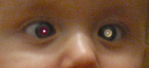
Differential of Leukocoria (White Pupil)
| Cause | Key clue | Laterality / setting |
|---|---|---|
| Retinoblastoma | Calcified retinal mass; family history | Uni- or bilateral; < 3 yr |
| Congenital cataract | Cloudy lens; TORCH, galactosemia | Newborn; uni/bilateral |
| Retinopathy of prematurity | Preterm + O₂; neovascularization | Bilateral; preterm |
| Coats disease | Retinal telangiectasia, exudates | Unilateral; boys |
| Persistent fetal vasculature (PHPV) | Microphthalmia (small eye) | Unilateral; term infant |
| Toxocariasis | Puppy exposure; retinal granuloma | Unilateral |

Genuine, high-yield associations (no invented acronyms):
- "Two hits, chromosome 13" — RB1 loss is the Knudson two-hit prototype; the same germline RB1 loss also predisposes to later osteosarcoma.
- "Bilateral = inherited" — bilateral/multifocal retinoblastoma is germline (hereditary) until proven otherwise; unilateral is usually sporadic.
- "Trilateral" = bilateral retinoblastoma + pinealoblastoma (midline intracranial "third eye").
- Patch the GOOD eye — the classic counter-intuitive rule for treating amblyopia.
- ROP = oxygen paradox — too much O₂ first, then relative hypoxia drives VEGF neovascularization.
Practice Ophthalmology now
Board-style questions, spaced-repetition flashcards, and a Socratic AI tutor — free to start.