Orbital & Preseptal Cellulitis
A Step 2 CK-focused lesson on distinguishing preseptal (anterior to the orbital septum, benign) from orbital (postseptal, vision- and life-threatening) cellulitis, driving the next-best-step decisions around CT imaging, IV antibiotics with MRSA coverage, and recognizing cavernous sinus thrombosis.
The orbital septum decides everything
The orbital septum — a fibrous sheet running from the orbital-rim periosteum (arcus marginalis) into the eyelid tarsal plates — is the anatomic line that names these two infections. Preseptal (periorbital) cellulitis stays anterior to the septum, involving only eyelid and periocular skin. Orbital (postseptal) cellulitis lies posterior to it, in the orbital fat and extraocular muscles. Both produce a red, warm, swollen, tender lid — but only orbital cellulitis threatens vision and life. The board's whole task is to separate them at the bedside: the septum-crossing signs — proptosis, painful or limited eye movements, and decreased vision — convert a benign-looking lid into an emergency. Orbital cellulitis most often arises from contiguous ethmoid sinusitis eroding through the paper-thin lamina papyracea (medial orbital wall), classically in a child.
- Preseptal: anterior to septum. Sources: lid trauma, insect bite, hordeolum/chalazion, dacryocystitis, impetigo. Bugs: S. aureus, S. pyogenes, S. pneumoniae.
- Orbital: posterior to septum. #1 source = bacterial (ethmoid) sinusitis via the lamina papyracea; can be polymicrobial — empirically cover MRSA.
- Postseptal red-flag signs: proptosis, pain with / limitation of EOM (ophthalmoplegia), diplopia, decreased acuity, RAPD, chemosis.
- Preseptal eye exam is normal: no proptosis, full painless movements, intact vision and pupils.
- Imaging of choice = CT of orbits and sinuses WITH contrast when orbital cellulitis is suspected → finds subperiosteal/orbital abscess.
- Orbital cellulitis = admit + IV antibiotics: vancomycin (for MRSA) plus ceftriaxone or ampicillin-sulbactam; add metronidazole for suspected intracranial/anaerobic spread; urgent ophthalmology + ENT consults.
- Surgical drainage for a drainable abscess, visual compromise, or failure to improve in 24–48 h.
Preseptal vs orbital at a glance
| Feature | Preseptal (periorbital) | Orbital (postseptal) |
|---|---|---|
| Location | Anterior to septum | Posterior to septum (orbital contents) |
| Typical source | Lid trauma, bite, hordeolum, dacryocystitis | Ethmoid sinusitis (lamina papyracea) |
| Proptosis | Absent | Present |
| Eye movements | Full, painless | Painful, limited (ophthalmoplegia) |
| Vision / pupil | Normal | May be decreased / RAPD |
| Chemosis, diplopia | Absent | Often present |
| Systemic illness | Mild | Fever, toxic |
| Imaging | Usually clinical | CT orbit + sinus w/ contrast |
| Treatment | Oral antibiotics, outpatient | Admit, IV antibiotics ± surgery |
Vignette: A 7-year-old with a week of nasal congestion and sinus pain now has fever, a swollen erythematous right eyelid, pain on looking laterally, a bulging eye, and double vision.
Diagnosis: Orbital (postseptal) cellulitis — extension of ethmoid sinusitis.
Next best step: Obtain CT of the orbits and sinuses with contrast (assess for subperiosteal/orbital abscess) and admit for IV broad-spectrum antibiotics — vancomycin plus ceftriaxone or ampicillin-sulbactam — with ophthalmology + ENT consultation. Start antibiotics emergently; do not delay treatment for imaging if vision is threatened.
Pearl: proptosis + painful or limited eye movement = orbital until proven otherwise.

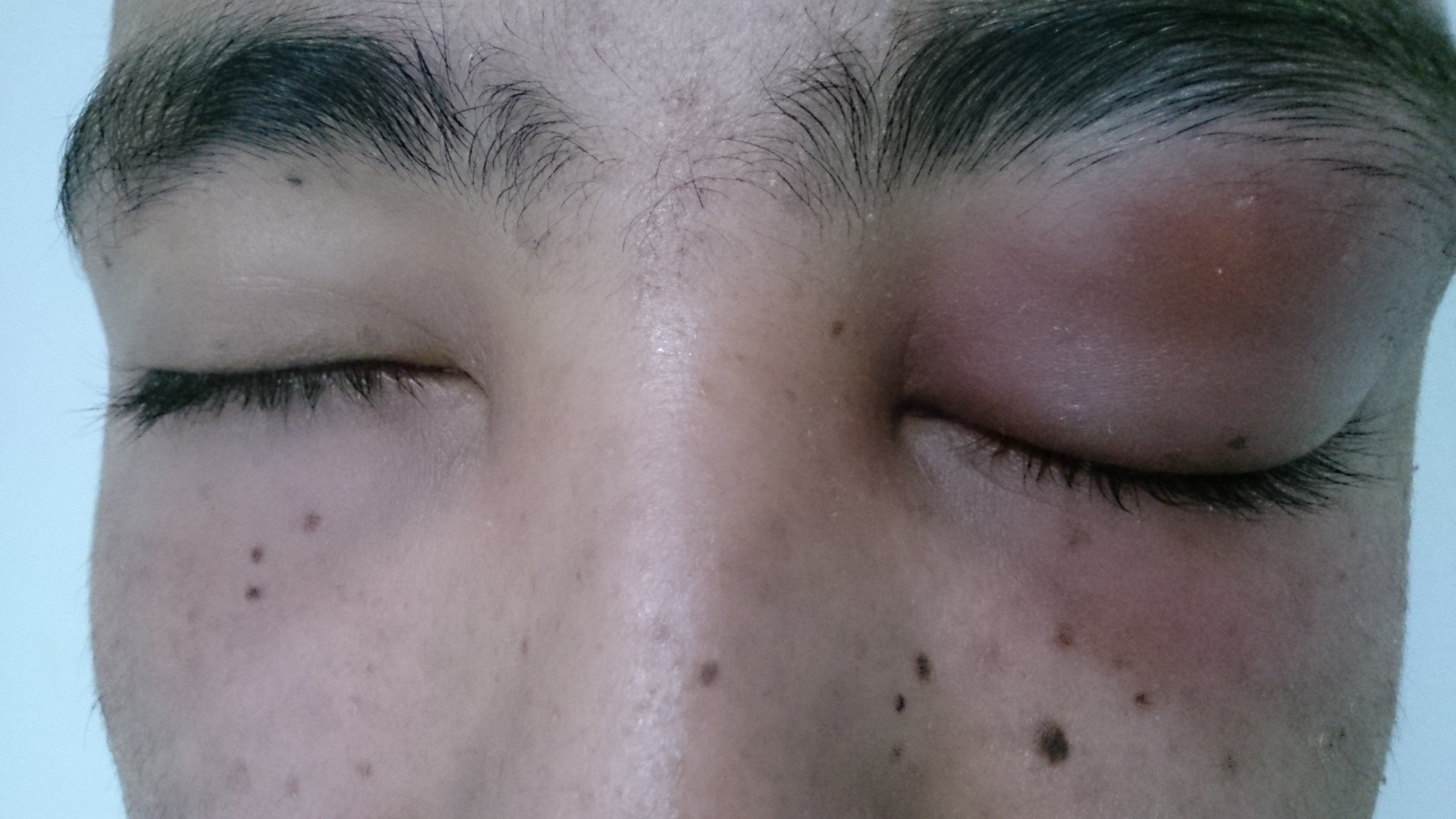
Vignette A: A 4-year-old has a red, swollen, tender left upper lid 2 days after an insect bite. Eye movements are full and painless, vision and pupils are normal, and there is no proptosis. → Preseptal cellulitis → oral antibiotics covering S. aureus (including MRSA) and streptococci — e.g., clindamycin alone, or TMP-SMX plus amoxicillin-clavulanate — with close follow-up; low threshold to image/admit if very young, toxic, or worsening.
Vignette B (danger): A patient with orbital cellulitis develops bilateral proptosis and chemosis, a CN VI palsy, and multiple cranial neuropathies. → Suspect cavernous sinus thrombosis → MRI / MR venography, continue IV antibiotics, and consider anticoagulation.
Cavernous sinus thrombosis (CST) is the feared complication — recall the sinus's contents with "O TOM CAT":
- O — Oculomotor (CN III)
- T — Trochlear (CN IV)
- O — Ophthalmic (CN V1)
- M — Maxillary (CN V2)
- C — internal Carotid artery
- A — Abducens (CN VI)
- T — sympathetic fibers (periarterial plexus)
Pearl: the internal carotid and CN VI run through the center of the sinus (III, IV, V1, V2 sit in the lateral wall), so abducens is often affected first → isolated lateral-gaze palsy. CST clues: bilateral eye findings, rapidly progressive proptosis/chemosis, multiple cranial-nerve palsies, headache, and a toxic-appearing patient.
- Complications of orbital cellulitis: subperiosteal abscess (most common), orbital abscess, vision loss (optic neuropathy, central retinal artery occlusion), cavernous sinus thrombosis, meningitis / intracranial abscess.
- Chandler classification (I–V): I = preseptal → II = orbital cellulitis → III = subperiosteal abscess → IV = orbital abscess → V = cavernous sinus thrombosis.
- Don't wait: any decreased acuity, RAPD, or proptosis is an ophthalmologic emergency — start IV antibiotics immediately.
- Key differential — invasive fungal (mucormycosis): a diabetic in DKA or immunocompromised patient with orbital signs plus a black necrotic eschar / turbinate needs emergent antifungals (amphotericin B) + surgical debridement, not just antibacterials.
Practice Ophthalmology now
Board-style questions, spaced-repetition flashcards, and a Socratic AI tutor — free to start.