Obstructive Sleep Apnea
A boards-focused walkthrough of obstructive sleep apnea from pharyngeal-collapse pathophysiology through polysomnography-based diagnosis and the CPAP-first management ladder, with STEP 2 CK next-best-step decisions, an OSA-vs-central comparison, and adult and pediatric vignettes.
Pathophysiology: airway collapse with preserved effort
Obstructive sleep apnea (OSA) is repetitive collapse of the pharyngeal airway during sleep, producing apneas (≥10 s cessation of airflow) and hypopneas despite continued respiratory effort — the feature that separates it from central sleep apnea. Each event drives hypoxemia (and often hypercapnia), triggering a cortical arousal that reopens the airway and fragments sleep. Chronic nocturnal hypoxemia plus repeated surges in sympathetic tone explain OSA's downstream damage: systemic (often resistant) hypertension, atrial fibrillation, pulmonary hypertension and cor pulmonale, stroke, and increased perioperative and motor-vehicle-accident risk. Anatomic and physiologic narrowing sets the stage — obesity with peripharyngeal fat, large neck circumference, retrognathia, macroglossia, tonsillar hypertrophy, and airway-relaxing agents (alcohol, sedatives). On boards, the sleepy, snoring, obese hypertensive man is OSA until proven otherwise.
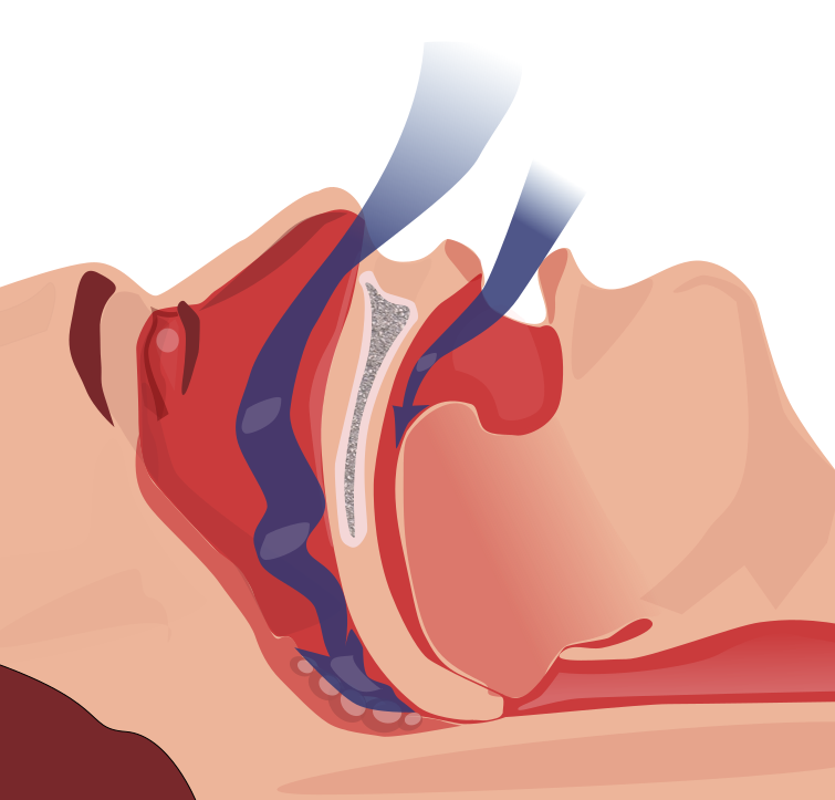
- Apnea = ≥10 s airflow cessation; hypopnea = reduced airflow + desaturation/arousal
- AHI (apnea–hypopnea index) = events/hour → mild 5–15, moderate 15–30, severe >30
- Diagnose OSA: AHI ≥5 with symptoms OR AHI ≥15 regardless of symptoms
- Risk factors: obesity, male sex, older age, neck >17 in (men)/16 in (women), retrognathia, tonsillar hypertrophy (children), alcohol/sedatives, hypothyroidism, acromegaly
- Nocturnal clues: loud snoring, witnessed apneas/gasping, nocturia, restless sleep
- Daytime: excessive somnolence (quantify with the Epworth scale), morning headache, poor concentration
- Complications: resistant HTN, AFib, pulmonary HTN/cor pulmonale, secondary erythrocytosis (↑Hct), motor-vehicle accidents
Diagnosis: test, don't assume
Diagnosis hinges on objective sleep testing, not symptoms alone. In-laboratory polysomnography (PSG) is the gold standard and the answer whenever the picture is complicated by significant cardiopulmonary disease, neuromuscular disease, suspected central apnea, or hypoventilation. Home sleep apnea testing (HSAT) is acceptable for an uncomplicated patient with high pretest probability of moderate-to-severe OSA; a negative or technically inadequate HSAT must be followed by PSG. Supportive labs are not diagnostic but are tested: chronic hypoxemia causes secondary erythrocytosis (↑Hgb/Hct), and a persistently elevated serum bicarbonate should prompt an ABG and suspicion for obesity hypoventilation syndrome (BMI ≥30 + daytime PaCO₂ ≥45 mmHg). Check TSH when hypothyroidism is plausible. Spirometry and CXR are typically normal in pure OSA and mainly serve to exclude mimics.
STOP-BANG — score ≥3 flags high risk; especially useful for preoperative screening:
- S — Snoring (loud)
- T — Tiredness / daytime sleepiness
- O — Observed apneas
- P — high blood Pressure
- B — BMI >35 kg/m²
- A — Age >50
- N — Neck circumference >40 cm
- G — Gender male
Vignette: A 52-year-old man with BMI 36 reports years of loud snoring; his wife has witnessed him stop breathing and gasp awake. He dozes off at red lights and while watching TV. BP is 152/96 on three antihypertensives. Exam: crowded oropharynx, neck 44 cm.
Most likely diagnosis: Obstructive sleep apnea — which also explains his resistant hypertension.
Best next step: Order polysomnography to confirm and grade severity — not empiric CPAP and not another BP drug first.
After confirmation: First-line therapy is CPAP plus weight loss and avoidance of alcohol/sedatives. Effective CPAP improves daytime sleepiness and can improve blood-pressure control.
Obstructive vs central sleep apnea
| Feature | Obstructive SA | Central SA |
|---|---|---|
| Mechanism | Airway collapse; effort present | ↓CNS drive; no effort |
| Airflow vs effort | Airflow absent despite chest/abdominal effort | Airflow and effort both absent |
| Classic causes | Obesity, large neck, tonsils | CHF (Cheyne–Stokes), opioids, stroke, high altitude |
| Snoring | Prominent | Often minimal |
| Body habitus | Obese | Variable / normal |
| First-line Rx | CPAP, weight loss | Treat cause; CPAP/ASV (avoid ASV if EF ≤45%) |
- Behavioral (all patients): weight loss (can be curative), avoid alcohol/sedatives/opioids, positional therapy (avoid supine sleep), treat nasal congestion
- CPAP = first-line and most effective; improves sleepiness, BP, and quality of life — adherence is the main limitation
- Mandibular advancement (oral) appliance: mild–moderate OSA or CPAP-intolerant
- Hypoglossal nerve stimulator: selected moderate–severe, CPAP-intolerant, lower-BMI patients (typically BMI <32–35)
- Surgery: UPPP in selected adults; adenotonsillectomy is FIRST-LINE in children
- Overlap/OHS: consider BiPAP when hypoventilation or coexisting COPD is present
- Counsel on drowsy driving; treating OSA lowers perioperative and cardiovascular risk

Vignette: A 5-year-old has loud snoring, restless sleep with witnessed pauses, chronic mouth-breathing, morning irritability, and declining school performance. Exam shows 4+ (kissing) tonsils and adenoidal facies; he is not obese.
Most likely diagnosis: Pediatric OSA from adenotonsillar hypertrophy — the leading cause in children (whereas obesity is the usual adult driver).
Best next step: Refer for adenotonsillectomy, the first-line treatment for childhood OSA. Polysomnography is the pediatric diagnostic gold standard and is obtained when the diagnosis is uncertain or the surgical benefit is unclear; CPAP is reserved for residual disease or poor surgical candidates.
Practice Pulmonary now
Board-style questions, spaced-repetition flashcards, and a Socratic AI tutor — free to start.