Nervous Tissue Histology
A Step 1–focused histology lesson on nervous tissue that maps neuron ultrastructure and axonal transport, then pairs each CNS/PNS glial cell with its origin, function, marker (GFAP, S-100), and disease. Reinforced with schwannoma and glioblastoma vignettes and classic transport/marker mnemonics.
Nervous tissue has two cell classes: neurons (excitable, conduct signals) and neuroglia (support cells that outnumber neurons and — unlike neurons — retain the ability to divide, which is why most primary CNS tumors are glial). Neurons are permanent (G0) cells with essentially no CNS regeneration. The dominant Step 1 theme is matching each glial cell to its origin, function, immunohistochemical marker, and disease. CNS glia: astrocytes, oligodendrocytes, microglia, ependymal cells — all neuroectoderm-derived except microglia, which are mesodermal (monocyte/macrophage lineage). PNS glia: Schwann cells and satellite cells (both neural crest). Two markers are tested relentlessly: GFAP (astrocytes) and S-100 (Schwann cells and other neural-crest derivatives).
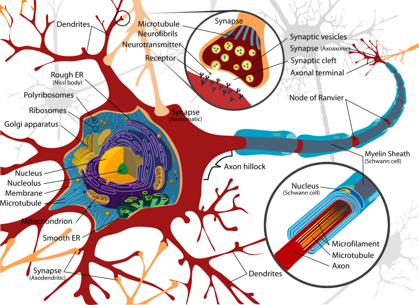
- Soma (perikaryon): large central nucleus with prominent nucleolus + Nissl substance = rough ER + free ribosomes (basophilic; site of protein / peptide-neurotransmitter synthesis)
- Nissl bodies are present in the soma and dendrites but NOT in the axon or axon hillock — the hillock is where the action potential is initiated
- Anterograde transport (soma → terminal) uses kinesin; retrograde transport (terminal → soma) uses dynein — both are ATP-dependent, microtubule-based motors
- Retrograde transport carries certain neurotropic agents toward the soma/CNS: rabies, HSV, tetanus toxin (poliovirus also uses this route)
- Chromatolysis = dispersal of Nissl substance + cell-body swelling + peripheral displacement of the nucleus after axonal injury (a reparative reaction)
- Morphology: multipolar (most CNS neurons), pseudounipolar (dorsal root / sensory ganglia), bipolar (retina, olfactory epithelium, CN VIII ganglia)
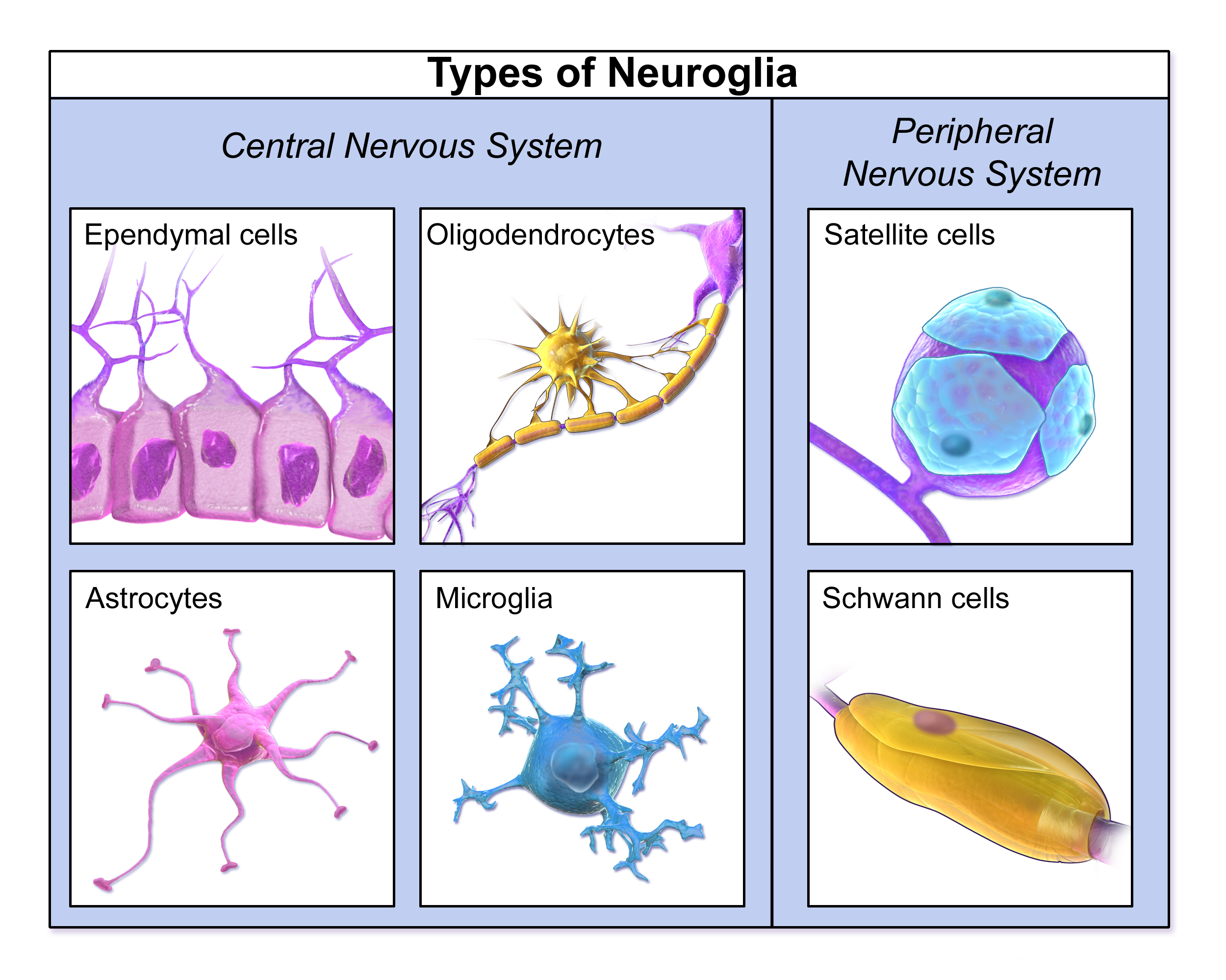
Glial cells: origin, function, marker, disease
| Glial cell | Location / origin | Key function | Marker & disease |
|---|---|---|---|
| Astrocyte | CNS / neuroectoderm | BBB support, K⁺ buffering, repair via gliosis | GFAP; astrocytoma / GBM |
| Oligodendrocyte | CNS / neuroectoderm | Myelinates many CNS axons | Injured in MS, PML; "fried-egg" oligodendroglioma |
| Microglia | CNS / mesoderm (monocyte lineage) | Phagocytic CNS macrophage | HIV → microglial nodules, multinucleated giant cells |
| Ependymal | CNS / neuroectoderm | Line ventricles / central canal, circulate CSF | Ependymoma |
| Schwann cell | PNS / neural crest | Myelinates one segment of one PNS axon; aids regeneration | S-100; schwannoma; injured in GBS |
| Satellite cell | PNS / neural crest | Surround & support ganglion cell bodies | Regulate ganglionic microenvironment |
- Myelin speeds conduction via saltatory conduction — the impulse jumps between nodes of Ranvier, bare axon segments with a high density of voltage-gated Na⁺ channels
- Oligodendrocyte: one cell sends multiple processes to myelinate many (up to ~30) CNS axons
- Schwann cell: one cell myelinates a single segment of a single PNS axon; also secretes factors that guide PNS axon regeneration
- CNS axons regenerate poorly — inhibitory astrocytic glial scar + no Schwann-cell guidance
- Wallerian degeneration: axon degenerates distal to a transection with myelin breakdown; the proximal stump can regrow in the PNS
- Demyelinating disease: central = MS, PML (JC virus) → oligodendrocytes; peripheral = Guillain-Barré, Charcot-Marie-Tooth → Schwann cells / myelin
- Connective-tissue sheaths of a peripheral nerve, inner → outer:
- Endoneurium — delicate CT around each individual axon (inflammatory target in Guillain-Barré)
- Perineurium — surrounds a nerve fascicle; forms the blood-nerve permeability barrier; the layer rejoined in microsurgical repair
- Epineurium — dense CT enclosing the whole nerve; carries the vasa nervorum
- Memory order: Endo → Peri → Epi (innermost to outermost)
- Dorsal root ganglia: pseudounipolar sensory neurons wrapped by satellite cells; cell bodies lie outside the CNS
- Autonomic ganglia: multipolar postganglionic cell bodies
- Gray matter = neuronal cell bodies + neuropil; white matter = myelinated axon tracts
Stem: A 46-year-old woman has progressive unilateral sensorineural hearing loss, tinnitus, and imbalance. MRI shows a contrast-enhancing mass at the cerebellopontine angle widening the internal acoustic meatus. Histology: spindle cells in alternating dense cellular zones (Antoni A, containing Verocay bodies) and loose myxoid zones (Antoni B); tumor is diffusely S-100 positive.
- Diagnosis: Vestibular schwannoma (acoustic neuroma) — a benign Schwann-cell tumor of the vestibular division of CN VIII
- Key branch point: bilateral vestibular schwannomas ⇒ Neurofibromatosis type 2 (NF2) — suspect strongly in a younger patient
- Next best step: MRI of the brain with gadolinium to confirm and size the lesion; definitive management is surgical resection or stereotactic radiosurgery, with observation for small / slow-growing tumors
Stem: A 61-year-old man has 8 weeks of worsening headaches and a new focal seizure. MRI shows a ring-enhancing mass crossing the corpus callosum ("butterfly glioma"). Biopsy: hypercellular, pleomorphic astrocytes with pseudopalisading necrosis and microvascular (endothelial) proliferation; tumor cells are GFAP-positive.
- Diagnosis: Glioblastoma (GBM), WHO grade 4 astrocytoma — the most common primary malignant brain tumor in adults
- Why GFAP matters: it confirms astrocytic (glial) origin, helping distinguish the tumor from metastasis or CNS lymphoma
- Management: maximal safe surgical resection followed by radiation + temozolomide; prognosis remains poor (median survival roughly 12–15 months)
- Kinesin = anterograde: "Kinesin Kicks it forward" — carries cargo away from the soma toward the axon terminal (microtubule + end)
- Dynein = retrograde: "Dynein Drags it back" — returns cargo to the soma (toward the microtubule − end)
- Retrograde neurotropic agents hitchhike toward the soma/CNS: rabies, HSV, tetanus toxin (poliovirus also)
- GFAP → Astrocytes (also marks astrocytomas / GBM); S-100 → Schwann cells and neural-crest tumors
- Antoni A + Verocay bodies = schwannoma; "fried-egg" cells = oligodendroglioma
Practice Histology now
Board-style questions, spaced-repetition flashcards, and a Socratic AI tutor — free to start.