Nephrotic Syndromes
A board-focused walkthrough of nephrotic syndrome: podocyte pathophysiology, the classic tetrad and its hypercoagulable and infectious complications, and the biopsy buzzwords, antibody tests, and next-best-step decisions that distinguish minimal change, FSGS, membranous, MPGN, diabetic, and amyloid disease.
Pathophysiology: it's a podocyte problem
Nephrotic syndrome is a glomerular disorder defined by massive proteinuria (>3.5 g/1.73 m²/day), hypoalbuminemia (<3.0–3.5 g/dL), edema, and hyperlipidemia with lipiduria. The lesion sits at the podocyte: injury causes foot-process effacement and loss of the size- and charge-selective filtration barrier, so albumin and larger proteins leak into the urine.
Contrast this with nephritic syndrome (an inflammatory picture: hematuria, RBC casts, hypertension, azotemia, and sub-nephrotic proteinuria). A few diseases — MPGN, diffuse proliferative lupus nephritis, and sometimes IgA nephropathy — can straddle both.
Every downstream consequence traces back to urinary protein loss: edema (low plasma oncotic pressure ± renal Na⁺ retention), a hypercoagulable state (urinary loss of antithrombin III), infection risk (loss of IgG/complement), and hyperlipidemia (hepatic lipoprotein synthesis rises and lipoprotein catabolism falls as oncotic pressure drops).
- Tetrad: proteinuria >3.5 g/day + hypoalbuminemia + edema + hyperlipidemia; frothy/foamy urine is the classic complaint.
- Urine sediment: oval fat bodies and fatty casts; under polarized light they form a "Maltese cross."
- Hypercoagulability: urinary loss of antithrombin III → renal vein thrombosis (most associated with membranous). Suspect it with sudden flank pain, gross hematuria, or a new PE.
- Infection: urinary IgG loss → encapsulated organisms; S. pneumoniae causing spontaneous bacterial peritonitis is classic in nephrotic children.
- Edema is typically periorbital in kids, dependent/pitting in adults.
- Epidemiology cheat sheet: kids → minimal change; adults → FSGS (esp. Black patients) and membranous (esp. White patients); diabetes is the #1 overall cause of nephrotic-range proteinuria.
Biopsy patterns — the core comparison
| Disease | Light microscopy | IF / EM | Buzzwords & associations |
|---|---|---|---|
| Minimal change | Normal | IF neg; EM: diffuse foot-process effacement | Kids #1; Hodgkin lymphoma, NSAIDs; steroid-responsive |
| FSGS | Focal & segmental sclerosis | IF: nonspecific IgM/C3; EM: effacement | Black patients, HIV, heroin, obesity, sickle cell; APOL1 |
| Membranous | Diffuse capillary-wall thickening; "spike and dome" (silver) | IF: granular IgG + C3; EM: subepithelial deposits | Anti-PLA2R (primary); SLE, HBV, solid tumors, NSAIDs/gold (secondary) |
| MPGN | GBM splitting, "tram-track" | IF: granular; EM: type I subendothelial, type II intramembranous (DDD) | HCV + cryoglobulins (I); C3 nephritic factor (II); nephritic–nephrotic overlap |
| Diabetic | Mesangial expansion, Kimmelstiel–Wilson nodules | EM: GBM thickening | #1 overall; hyperfiltration early |
| Amyloid | Congo red → apple-green birefringence | EM: nonbranching fibrils | AL (myeloma), AA (chronic inflammation) |
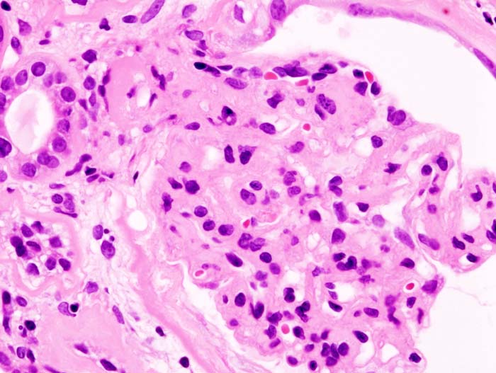
Vignette: A 4-year-old boy develops periorbital edema and frothy urine about a week after an upper-respiratory infection. BP is normal. UA: 4+ protein, no blood, bland sediment (no RBC casts). Labs: albumin 2.0 g/dL, elevated cholesterol.
Diagnosis: Minimal change disease — the most common nephrotic syndrome in children. Classically selective (albumin) proteinuria with a normal-appearing glomerulus on light microscopy and foot-process effacement on EM.
Next best step: Start empiric oral corticosteroids (prednisone). A typical child does not need a biopsy first because MCD is highly steroid-responsive. Reserve renal biopsy for steroid-resistant, frequently relapsing, or atypical cases (age <1 or >12, hematuria, hypertension, renal insufficiency, or low complement).
Vignette: A 55-year-old man has weeks of lower-extremity edema and foamy urine; a 24-hour collection shows 6 g of protein and albumin is low. He then develops acute left flank pain with gross hematuria and worsening leg swelling.
Diagnosis: Membranous nephropathy complicated by renal vein thrombosis — the classic hypercoagulable complication (urinary antithrombin III loss). Biopsy would show subepithelial "spike-and-dome" deposits with granular IgG/C3 on IF.
Next best steps:
- Check anti-PLA2R antibody (positive in ~70% of primary disease).
- Work up secondary causes: HBV/HCV, SLE (ANA), and age-appropriate malignancy screening (solid tumors).
- Anticoagulate the thrombosis; confirm with CT/MR venography or Doppler ultrasound.
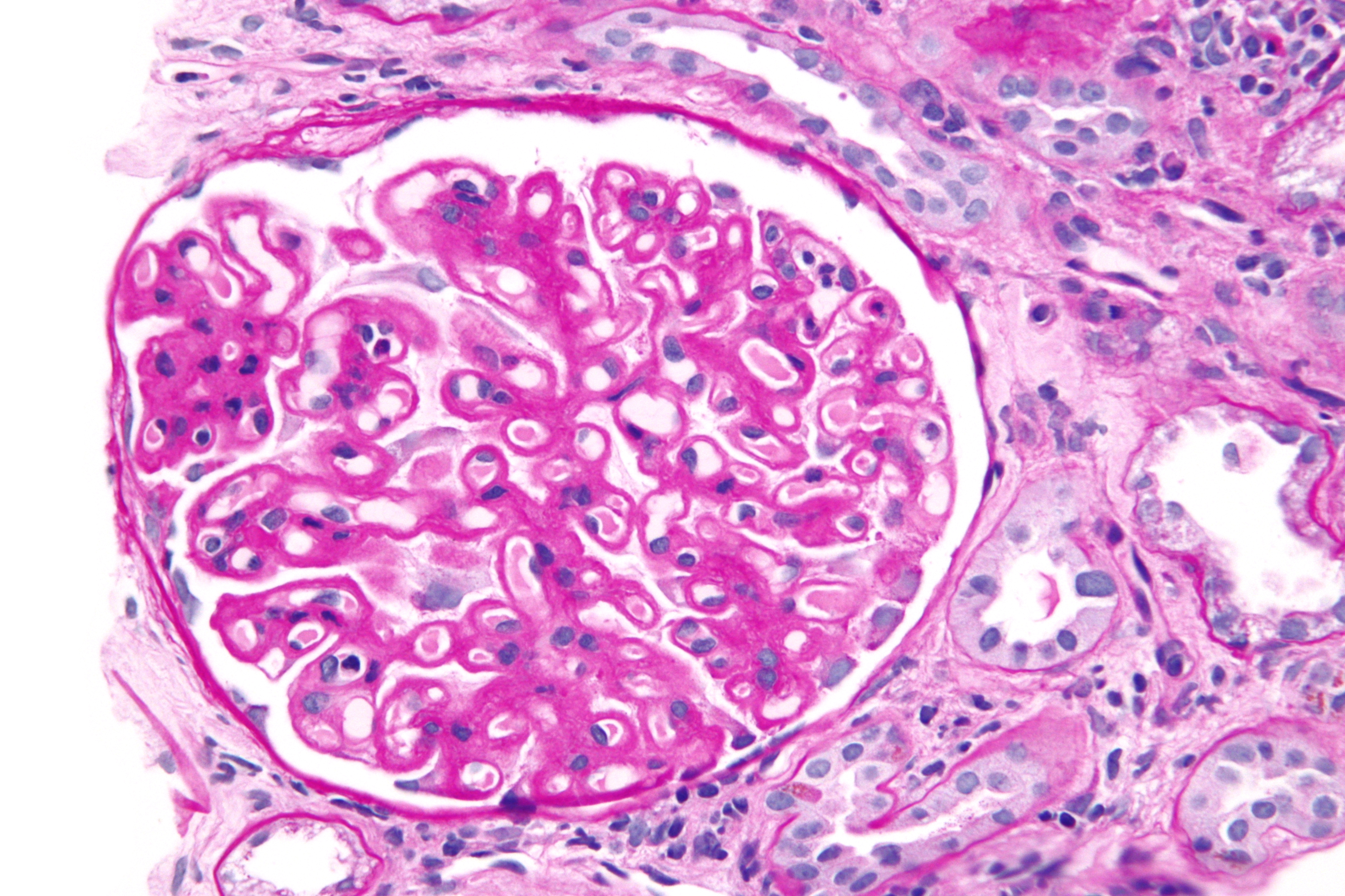
These are genuine board classics — see the phrase, name the disease:
- "Spike and dome" (silver stain, subepithelial) → Membranous
- "Tram-track" (GBM splitting) → MPGN
- "Kimmelstiel–Wilson nodules" → Diabetic nephropathy
- "Apple-green birefringence" (Congo red, polarized) → Amyloidosis
- Normal LM + foot-process effacement on EM → Minimal change
- "Maltese cross" (polarized urine) → lipiduria / oval fat bodies of any nephrotic syndrome
Antibody pearls: anti-PLA2R = primary membranous; C3 nephritic factor = MPGN type II (dense deposit disease).
Confirm & quantify proteinuria
- Spot urine protein:creatinine (or albumin:creatinine) ratio estimates g/day; nephrotic range is ≥3–3.5. A 24-hour collection is the reference standard.
- UA + microscopy: heavy protein, fatty casts, oval fat bodies, Maltese crosses; bland (non-nephritic) sediment.
- Serum: low albumin, high lipids; check renal function.
Find the cause (adults)
- Screen for diabetes (most common), HBV/HCV, HIV, SLE (ANA), SPEP/UPEP + serum free light chains (myeloma/amyloid), and anti-PLA2R (membranous).
Biopsy
- Indicated in most adults to establish the diagnosis and guide therapy; often deferred in children with a typical minimal-change presentation (treat empirically first).
General (all nephrotic patients)
- ACE inhibitor or ARB to lower intraglomerular pressure and proteinuria (plus BP control).
- Loop diuretic + sodium restriction for edema.
- Statin for hyperlipidemia; treat/withdraw the underlying cause (glycemic control, stop offending drug, treat infection/malignancy).
- Consider anticoagulation in high-risk membranous (very low albumin).
Disease-specific
- Minimal change: corticosteroids (excellent response, esp. kids); relapses → steroid-sparing agents (calcineurin inhibitors, cyclophosphamide).
- FSGS: ACEi/ARB + corticosteroids; less responsive; may recur after transplant.
- Membranous: supportive if low-risk; rituximab or cyclophosphamide-based (Ponticelli) regimens if high-risk/progressive.
- Diabetic: glycemic + BP control, ACEi/ARB, SGLT2 inhibitors.
Practice Renal now
Board-style questions, spaced-repetition flashcards, and a Socratic AI tutor — free to start.