Nephrolithiasis (Kidney Stones)
A boards-focused walkthrough of nephrolithiasis — stone chemistry with classic crystal buzzwords, renal-colic presentation, CT-first diagnosis, and size-based plus emergency management — anchored on the high-yield infected-obstructed-stone next-best-step decision.
Pathophysiology: Why Stones Form
Nephrolithiasis occurs when urinary solute concentration exceeds solubility, driving crystal nucleation, aggregation, and growth. The balance tips toward stones with low urine volume (dehydration — the single biggest modifiable risk), supersaturation of stone-forming solutes (calcium, oxalate, uric acid, cystine), and loss of inhibitors (low urinary citrate, low magnesium). Urine pH is decisive: acidic urine favors uric acid and cystine; alkaline urine favors calcium phosphate and struvite. About 80% of stones are calcium-based (mostly calcium oxalate). Stones become symptomatic when they migrate into the ureter and obstruct, raising intraluminal and renal-pelvic pressure; prostaglandins drive the ureteral smooth-muscle response and renal colic — which is the rationale for NSAIDs. Boards test the metabolic setup (who forms which stone), the buzzword crystal shape, the imaging choice, and the next best step — above all, recognizing the infected, obstructed stone that demands emergent drainage.
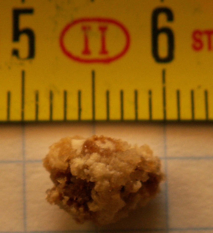
- Calcium oxalate (~75-80%, most common): radiopaque; envelope / dumbbell crystals; acidic-to-neutral urine. Causes: idiopathic hypercalciuria, hyperoxaluria (enteric — Crohn / gastric bypass / short bowel), hypocitraturia, ethylene glycol, vitamin C excess.
- Calcium phosphate (~5-10%): radiopaque; alkaline urine; distal (type 1) RTA, primary hyperparathyroidism, carbonic anhydrase inhibitors (topiramate, acetazolamide).
- Struvite (Mg-ammonium-phosphate, ~10-15%): staghorn calculi; coffin-lid crystals; alkaline urine from urease-positive bugs (Proteus, Klebsiella, Staph saprophyticus).
- Uric acid (~5-10%): radiolucent (CT only — invisible on plain film); acidic urine; rhomboid / rosette crystals; gout, high cell turnover (tumor lysis, leukemia), obesity / diabetes.
- Cystine (~1%): young patient + family history; hexagonal crystals; autosomal-recessive cystinuria; positive urinary nitroprusside (cyanide) test; can form staghorns.
Stone Comparison Table
| Stone | Approx % | X-ray | Urine pH | Crystal shape | Key associations |
|---|---|---|---|---|---|
| Calcium oxalate | 75-80 | Radiopaque | Acidic/normal | Envelope / dumbbell | Hypercalciuria; hyperoxaluria (Crohn, bypass); low citrate |
| Calcium phosphate | 5-10 | Radiopaque | Alkaline | Wedge-shaped / amorphous | Distal (type 1) RTA; hyperparathyroidism; topiramate |
| Struvite | 10-15 | Radiopaque | Alkaline | Coffin-lid | Urease+ (Proteus); staghorn |
| Uric acid | 5-10 | Radiolucent | Acidic | Rhomboid / rosette | Gout; tumor lysis; obesity/DM |
| Cystine | ~1 | Faintly opaque | Acidic | Hexagonal | Cystinuria (AR); children + FHx |
Presentation
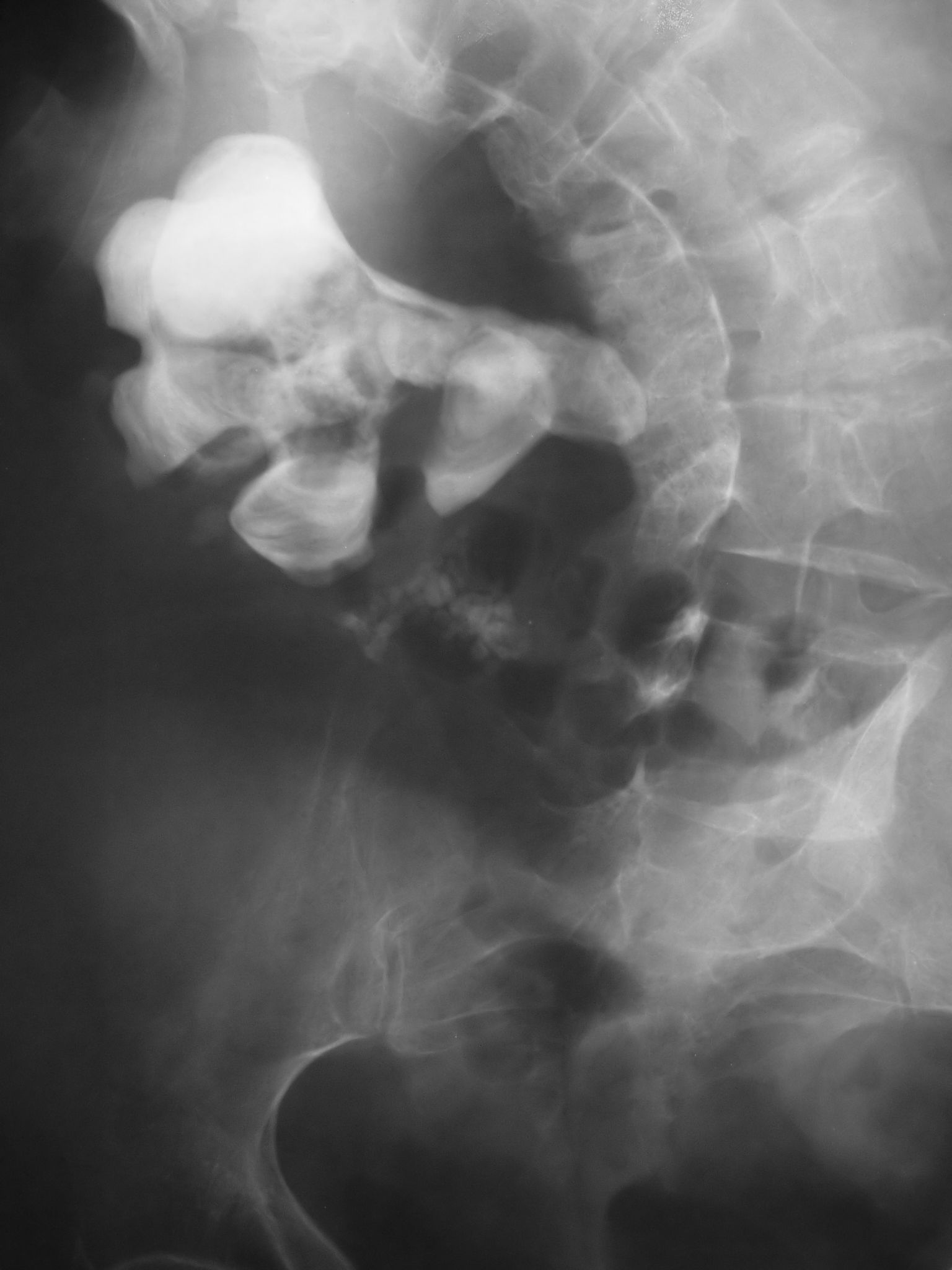
- Renal colic: acute, severe, colicky flank pain radiating to the groin; patient writhing, cannot lie still (contrast peritonitis — lies motionless).
- Hematuria (gross or micro), nausea/vomiting; pain migrates flank → groin → dysuria/frequency as the stone nears the bladder.
- 3 sites of impaction: UPJ, pelvic brim (ureter crosses the iliac vessels), UVJ (narrowest, most common site to lodge).
Diagnosis / next best step
- Non-contrast helical CT (CT KUB) = gold standard, most sensitive and specific; detects even radiolucent uric acid stones.
- Ultrasound first-line in pregnancy and children (no radiation); may miss small or mid-ureteral stones.
- Urinalysis: hematuria, crystals, pH (<5.5 → uric acid; >7 → struvite / Ca-phosphate); obtain urine culture.
- Labs: BMP/creatinine, calcium, uric acid. Recurrent stones → 24-hour urine (volume, Ca, oxalate, citrate, uric acid, pH) plus stone composition analysis.

Vignette: A 58-year-old woman has 2 days of right flank pain, now with fever 39.2°C, rigors, and hypotension. Exam: right CVA tenderness. Labs: WBC 18,000, rising creatinine; UA shows pyuria, positive nitrites, and alkaline pH. Non-contrast CT: a 9-mm obstructing right ureteral stone with hydronephrosis.
Diagnosis: Obstruction + UTI = infected, obstructed kidney (impending urosepsis) — a urologic emergency and the single highest-yield next-best-step on boards.
Next best step: Emergent decompression with a retrograde ureteral stent or percutaneous nephrostomy, plus IV fluids and broad-spectrum antibiotics. Do not attempt definitive stone removal (lithotripsy / ureteroscopy) during active infection — drain first, treat the sepsis, then remove the stone electively later. The alkaline pH with pyuria and nitrites signals a urease-producing organism (e.g., Proteus) and raises suspicion for a struvite stone — but any obstructing stone can become secondarily infected, so composition is confirmed later by stone analysis and does not change the emergent drainage.
Vignette: A 55-year-old man with gout, obesity, and type 2 diabetes has acute left flank pain and gross hematuria. KUB shows no stone, yet non-contrast CT reveals a 6-mm left ureteral calculus. Urine pH is 5.0 with rhomboid crystals.
Diagnosis: Uric acid stone — the classic radiolucent stone (invisible on plain film, visible on CT) forming in persistently acidic urine.
Next best step: Hydration, analgesia, and medical dissolution via urinary alkalinization with potassium citrate (target urine pH ~6.5-7); add allopurinol if hyperuricemic/hyperuricosuric. Uric acid stones are among the few that can be dissolved medically — alkalinize rather than rushing to surgery. Add tamsulosin (medical expulsive therapy) to aid passage of the ureteral stone.
Acute
- NSAIDs (e.g., ketorolac) = first-line analgesia — as good as or better than opioids with less nausea; opioids are adjuncts. Add antiemetics and hydration.
- Medical expulsive therapy (MET): tamsulosin (alpha-blocker), most useful for distal ureteral stones ~5-10 mm.
By stone size (likelihood of spontaneous passage)
- <5 mm: usually passes → conservative, strain urine.
- 5-10 mm: trial of passage + MET.
- ~10-20 mm: shock-wave lithotripsy (ESWL) or ureteroscopy.
- >20 mm or staghorn: percutaneous nephrolithotomy (PCNL).
Emergent intervention (drain now): infection + obstruction, AKI or solitary/transplant kidney, or intractable pain/vomiting.
- COLA — dibasic amino acids lost in cystinuria: Cystine, Ornithine, Lysine, Arginine (only cystine is insoluble and forms stones).
- Coffin-lid crystals + staghorn + Proteus + alkaline urine → Struvite.
- Crystal shapes: Envelope → Ca oxalate; Hexagon → Cystine; Rhombus / rosette → Uric acid.
- Radiolucent on X-ray but visible on CT → Uric acid.
- Patient writhing, cannot stay still → renal colic (vs. peritonitis, where the patient lies perfectly still).
- Fluids first: intake to produce >2-2.5 L of urine/day — the single most effective prevention for all stone types.
- Calcium oxalate: thiazide (lowers urinary calcium), potassium citrate (raises citrate), low sodium, moderate animal protein, reduce dietary oxalate — but keep normal dietary calcium (low-calcium diets paradoxically increase oxalate absorption and stone risk).
- Uric acid: potassium citrate alkalinization ± allopurinol; low-purine diet.
- Cystine: aggressive hydration + alkalinization; tiopronin / penicillamine if refractory.
- Struvite: complete surgical stone clearance + treat/eradicate infection; acetohydroxamic acid (urease inhibitor) in select refractory cases.
Practice Renal now
Board-style questions, spaced-repetition flashcards, and a Socratic AI tutor — free to start.