Neonatal Jaundice & Hyperbilirubinemia
A Step 2 CK–focused walkthrough of neonatal jaundice that separates benign physiologic and feeding-related unconjugated patterns from dangerous hemolytic and conjugated (biliary atresia) causes, anchoring on the two boards forks — bilirubin fraction and timing — and the exact next-best-step in both workup and management.
The Big Picture
Neonatal jaundice is yellow discoloration of the skin and sclera from bilirubin, clinically visible once total serum bilirubin (TSB) exceeds ~5 mg/dL. It affects roughly 60% of term and 80% of preterm infants. Newborns are primed for hyperbilirubinemia by three forces: a high red-cell mass with shorter RBC lifespan (↑ heme turnover), an immature hepatic UGT1A1 conjugating enzyme, and increased enterohepatic circulation.
Two forks decide every board question. Fork 1 — bilirubin fraction: unconjugated (indirect) bilirubin is fat-soluble, albumin-bound, and neurotoxic (crosses the blood–brain barrier → kernicterus), whereas conjugated (direct) bilirubin is water-soluble and in a neonate is ALWAYS pathologic. Fork 2 — timing: true physiologic jaundice never appears in the first 24 hours of life.
- Physiologic jaundice: appears after 24 h, peaks day 3–5 (term), always unconjugated, resolves by 1–2 weeks; peak TSB is typically ~12 mg/dL and, by definition, stays below the phototherapy threshold.
- Jaundice in the first 24 hours of life is ALWAYS pathologic — think hemolysis (ABO/Rh isoimmunization, G6PD deficiency) until proven otherwise.
- Other red flags: TSB rising > 0.2 mg/dL/hr or > 5 mg/dL/day, TSB > 95th percentile on the hour-specific (Bhutani) nomogram, or any direct/conjugated bilirubin > 1 mg/dL.
- Prolonged jaundice (> 2 weeks term, > 3 weeks preterm) → fractionate the bilirubin: if direct is elevated, work up biliary atresia; if indirect, consider breast-milk jaundice, hypothyroidism, or G6PD deficiency.
- Screen every newborn with transcutaneous bilirubin; confirm any elevated value with serum TSB — visual estimation of severity is unreliable and should not guide treatment.
Three Benign Unconjugated Patterns
| Feature | Physiologic | Breastfeeding-failure (early) | Breast-milk jaundice (late) |
|---|---|---|---|
| Onset / peak | After 24 h; peaks day 3–5 | Day 2–4 (first week) | Starts day 5–7; peaks ~2 wk; can last weeks |
| Mechanism | Immature UGT + ↑ enterohepatic circulation | Poor intake → dehydration, ↓ stooling → ↑ enterohepatic circulation | Factors in breast milk (classically β-glucuronidase) ↑ enterohepatic reabsorption |
| Baby's status | Well | Dehydrated, > 10% weight loss, few wet diapers | Thriving, gaining weight |
| Management | Observe | Increase feeding frequency + lactation support (do NOT routinely stop breastfeeding) | Continue breastfeeding; benign, self-limited |
Buzzwords: Term newborn, mother blood type O-positive, becomes visibly jaundiced at 18 hours of life; elevated TSB, pallor, hepatosplenomegaly.
Jaundice before 24 h = hemolysis until proven otherwise (isoimmune > enzymatic).
NEXT BEST STEP — workup:
- TSB with direct fraction, CBC with peripheral smear, reticulocyte count.
- Infant blood type + Rh and direct antiglobulin (Coombs) test; check mother's type and antibody screen.
- Interpret: mother O + infant A or B with positive DAT → ABO hemolytic disease. Smear = spherocytes (ABO/hereditary spherocytosis) vs bite cells / Heinz bodies (G6PD deficiency).
NEXT BEST STEP — management: start phototherapy per the hour-specific nomogram; consider IVIG if isoimmune and TSB keeps rising despite intensive phototherapy or nears the exchange level; escalate to exchange transfusion if TSB crosses the exchange threshold or any sign of acute bilirubin encephalopathy.
ABO vs. Rh Hemolytic Disease
| Feature | ABO incompatibility | Rh(D) isoimmunization |
|---|---|---|
| Setup | Mother O, infant A or B | Mother Rh-negative, infant Rh-positive |
| First pregnancy affected? | Yes (preformed maternal anti-A/anti-B IgG) | No — requires prior sensitization |
| Severity | Usually mild, gradual | Severe; can cause hydrops fetalis, marked anemia |
| Direct Coombs (DAT) | Weakly + / variable | Strongly positive |
| Smear | Spherocytes | Nucleated RBCs, polychromasia |
| Prevention | None | Anti-D immunoglobulin (RhoGAM) at 28 wk + within 72 h postpartum |
Buzzwords: 5-week-old with persistent jaundice, pale / clay-colored (acholic) stools, dark urine, and hepatomegaly; labs show elevated DIRECT (conjugated) bilirubin.
Conjugated hyperbilirubinemia is never physiologic — the must-not-miss diagnosis is biliary atresia (the leading indication for pediatric liver transplant).
NEXT BEST STEP — workup:
- Fractionate bilirubin (confirm the direct elevation).
- Abdominal ultrasound — absent/contracted gallbladder, "triangular cord sign."
- Hepatobiliary (HIDA) scan — liver takes up tracer but shows NO excretion into the bowel.
- Liver biopsy (bile-duct proliferation) → confirm with intraoperative cholangiogram (gold standard).
Treatment: Kasai hepatoportoenterostomy, ideally before 8 weeks (60 days) of life for best outcome; supplement fat-soluble vitamins A, D, E, K. Also exclude sepsis/TORCH, galactosemia, hypothyroidism, and choledochal cyst.
- Phototherapy = first-line. Blue–green light (~460–490 nm) converts skin unconjugated bilirubin to water-soluble lumirubin and photoisomers, excreted in bile and urine without hepatic conjugation. Thresholds come from the AAP hour-specific nomogram (gestational age + neurotoxicity risk factors), not a single magic number.
- Neurotoxicity risk factors that lower the treatment threshold: GA < 38 wk, isoimmune hemolytic disease, G6PD deficiency, sepsis, low albumin, clinical instability.
- IVIG may be considered for isoimmune hemolytic disease when TSB rises despite intensive phototherapy or nears the exchange level.
- Exchange transfusion (double-volume) for TSB above the exchange threshold or ANY sign of acute bilirubin encephalopathy — it removes bilirubin and antibody-coated RBCs.
- Optimize feeding/hydration and treat the underlying cause.
- Conjugated hyperbilirubinemia does NOT respond to phototherapy — using it in cholestasis can cause "bronze baby syndrome."
- Free unconjugated bilirubin crosses the BBB and deposits in the basal ganglia (globus pallidus), subthalamic nuclei, and brainstem — conjugated bilirubin does NOT cause kernicterus.
- Acute bilirubin encephalopathy progresses in stages: early lethargy, hypotonia, poor feeding → hypertonia with retrocollis / opisthotonos, high-pitched cry, fever → coma.
- Chronic kernicterus tetrad: (1) choreoathetoid cerebral palsy / dystonia, (2) sensorineural hearing loss (auditory neuropathy), (3) impaired upward gaze, (4) dental enamel dysplasia.
- Preterm and hypoalbuminemic infants are most vulnerable; sulfa drugs and ceftriaxone (albumin-displacers) raise risk — ceftriaxone is contraindicated in neonates.

- "Direct is never right" — any conjugated/direct hyperbilirubinemia in a neonate is pathologic → biliary atresia until proven otherwise.
- "Day 1 = hemolysis" — jaundice in the first 24 hours is never physiologic.
- "Starve early, thrive late" — breastfeeding-failure jaundice = early, dehydrated, underfed baby; breast-milk jaundice = late, thriving baby.
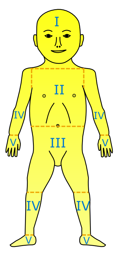
- Kramer's cephalocaudal rule — jaundice advances head → toe as bilirubin climbs (face ≈ 5, trunk ≈ 10, palms & soles > 15 mg/dL). A rough bedside clue only — always confirm with a measured level.
- Kernicterus lands in the basal ganglia → remember the triad of movement disorder (choreoathetosis) + hearing loss + upgaze palsy.
Practice Pediatrics now
Board-style questions, spaced-repetition flashcards, and a Socratic AI tutor — free to start.