Muscle Tissue Histology
A board-focused walkthrough of the three muscle types — how to identify skeletal, cardiac, and smooth muscle on histology, the sarcomere band anatomy and what changes during contraction, and the calcium-handling and intercalated-disc details Step 1 loves to test.
Why the boards test muscle histology
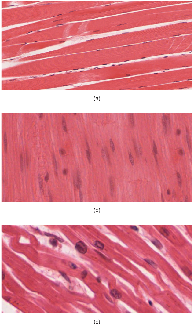
Muscle tissue comes in three flavors the boards make you distinguish on a slide or EM: skeletal, cardiac, and smooth. Step 1 rarely asks you to name a disease here — it asks you to identify the tissue from morphology, then reason about the mechanism (excitation–contraction coupling, calcium source, regeneration).
Nail three questions on any image:
- Striations present?
- How many nuclei, and where?
- Branching fibers with intercalated discs?
Those three features unambiguously separate all three types. Layered on top are the sarcomere ultrastructure (which bands shorten during contraction) and the calcium-handling differences that explain why cardiac muscle — but not skeletal — needs extracellular calcium to contract.
- Skeletal: striated; long, cylindrical, unbranched fibers; multiple peripheral nuclei; voluntary; regenerates via satellite cells (limited).
- Cardiac: striated; branching fibers; 1–2 central nuclei; joined by intercalated discs; involuntary; essentially no regeneration (heals by scar).
- Smooth: no striations; spindle/fusiform cells; single central nucleus; involuntary; regenerates well.
- Striations = sarcomeres in register. Skeletal and cardiac are striated; smooth is not (actin/myosin present but not in ordered sarcomeres).
- Peripheral nuclei → skeletal; central nuclei → cardiac. Central nuclei in adult skeletal fibers suggest regeneration or myopathy.
- Intercalated discs are unique to cardiac and contain gap junctions (electrical coupling → functional syncytium), fascia adherens (anchor actin), and desmosomes (macula adherens → anchor desmin intermediate filaments).
Skeletal vs cardiac vs smooth
| Feature | Skeletal | Cardiac | Smooth |
|---|---|---|---|
| Striations | Yes | Yes | No |
| Nuclei | Many, peripheral | 1–2, central | 1, central |
| Cell shape | Long cylindrical | Branching | Spindle (fusiform) |
| Intercalated discs | No | Yes | No |
| T-tubule unit | Triad (A–I junction) | Dyad (Z line) | None (has caveolae) |
| Ca²⁺ source | SR only | SR + extracellular | SR + extracellular |
| Ca²⁺ receptor protein | Troponin C | Troponin C | Calmodulin (no troponin) |
| Regeneration | Satellite cells (limited) | Minimal/none | Good |
| Control | Voluntary | Involuntary | Involuntary |
Sarcomere = Z line to Z line (the contractile unit).
- Z line (Z disc): anchors thin (actin) filaments; contains α-actinin.
- I band: light (Isotropic); thin filaments only; bisected by the Z line. Shortens.
- A band: dark (Anisotropic); spans the full thick (myosin) filament including overlap. Constant length.
- H zone: center of A band; thick filaments only (no overlap). Shortens.
- M line: center of H zone; anchors thick filaments (myomesin).
- Titin: Z line → M line; elastic recoil, prevents overstretch.
During contraction: the I band and H zone narrow; the A band stays the same. This is the sliding-filament principle — filaments slide past each other, they do not shorten.

Vignette: EM of myocardium shows fibers joined end-to-end by dense, step-like junctions containing gap junctions, fascia adherens, and desmosomes. A young athlete with a mutation weakening the desmosomal components of these junctions has ventricular arrhythmias and fibrofatty replacement of the RV.
- Structure shown → intercalated disc (found only in cardiac muscle).
- Gap junctions (connexons) give low-resistance electrical coupling → ventricles act as a functional syncytium (coordinated contraction).
- Diagnosis → arrhythmogenic right ventricular cardiomyopathy (ARVC), from desmosomal mutations (classically plakophilin-2) → arrhythmia + sudden death in young athletes.
- Next-step buzz: desmosomes anchor intermediate filaments (desmin); fascia adherens anchor actin; gap junctions pass ions/current.
How the action potential reaches the SR:
- Skeletal: AP down T-tubule → DHP receptor (voltage sensor) is mechanically coupled to RyR1 on the SR → SR Ca²⁺ release. No extracellular Ca²⁺ required.
- Cardiac: AP → L-type Ca²⁺ channel (DHP receptor) opens → extracellular Ca²⁺ enters → triggers RyR2 = calcium-induced calcium release (CICR). Requires extracellular Ca²⁺.
- Smooth: Ca²⁺ (extracellular + SR) binds calmodulin → activates MLCK → phosphorylates myosin light chain → contraction. No troponin. Relaxation via MLC phosphatase; sustained tone = latch state.
T-tubule anatomy: skeletal triad (T-tubule + 2 SR terminal cisternae) at the A–I junction; cardiac dyad (T-tubule + 1 SR cisterna) at the Z line.
- A band = Always the same length during contraction (dark, Anisotropic).
- I band = Isotropic, light, and thin — like the letter I, it holds the thin (actin) filaments; it shrinks.
- Which parts shorten: the H zone, I band, and the Z–Z (sarcomere) length all narrow during contraction — the A band does not.
- Nuclei: Peripheral → skeletal (Perimeter); Central → Cardiac.
- Calcium: skeletal = SR only (Self-sufficient); cardiac needs the Circulation (extracellular Ca²⁺ for CICR).
Vignette: A biopsy of injured skeletal muscle shows scattered small fibers with centrally located nuclei among mature fibers that have peripheral nuclei. Which cell mediated the repair?
- Answer → satellite cells: quiescent myogenic progenitors lying between the sarcolemma and the basal (external) lamina; they activate after injury to regenerate fibers.
- Pearl: central nuclei in adult skeletal muscle = regenerating (or myopathic) fibers. Mature skeletal nuclei are peripheral — don't misread this as cardiac muscle (normally central-nucleated).
- Contrast: cardiac myocytes lack an effective satellite-cell pool → infarcted myocardium heals by fibrous scar, not new muscle. Smooth muscle regenerates readily.
Practice Histology now
Board-style questions, spaced-repetition flashcards, and a Socratic AI tutor — free to start.