Multiple Sclerosis & Demyelinating Disease
A high-yield STEP 1/CK lesson on multiple sclerosis and the CNS demyelinating differential — pathophysiology, localization buzzwords (optic neuritis, INO, Lhermitte, Uhthoff), McDonald/MRI/CSF diagnosis, and next-best-step management including the classic NMOSD interferon trap.
Pathophysiology & Epidemiology
Multiple sclerosis (MS) is a chronic, immune-mediated demyelinating disease of the CNS. Autoreactive CD4-positive (Th1/Th17) and CD8-positive T cells plus B cells breach the blood–brain barrier and attack oligodendrocyte myelin, producing perivascular inflammation and demyelinated plaques with relative early axonal sparing; cumulative axonal loss later drives fixed disability.
Classic demographics: women aged 20–40 (F:M about 3:1), with rising prevalence at higher latitudes. Established risk factors: EBV infection (now viewed as a near-necessary trigger), low vitamin D, smoking, and HLA-DRB1*15:01 (HLA-DR2). Plaques favor periventricular white matter, optic nerve, brainstem/MLF, juxtacortical regions, and spinal cord — the anatomy behind every classic syndrome.
Course is most often relapsing–remitting (RRMS, ~85%), which may convert to secondary progressive; primary progressive MS (~10–15%) presents later, affects men relatively more (F:M nearer 1:1), and manifests as a slowly worsening myelopathy.

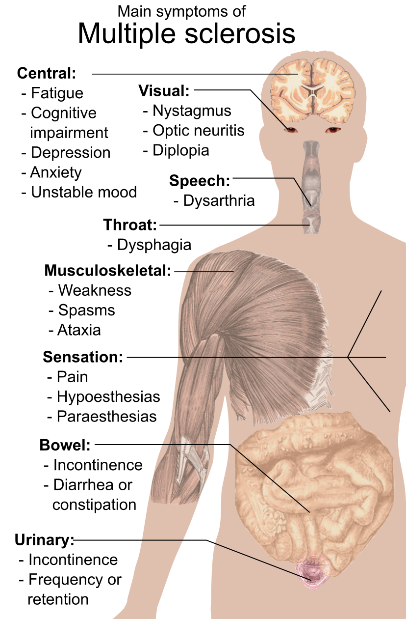
- Optic neuritis (common first attack): painful monocular vision loss, red-color desaturation, central scotoma, relative afferent pupillary defect (Marcus Gunn pupil)
- Internuclear ophthalmoplegia (INO): MLF lesion causes impaired adduction ipsilateral to the lesion plus nystagmus of the contralateral abducting eye; bilateral INO in a young adult is MS until proven otherwise
- Lhermitte sign: electric shock down the spine on neck flexion (dorsal columns)
- Uhthoff phenomenon: transient worsening with heat, exercise, or fever
- Charcot neurologic triad: scanning speech, intention tremor, nystagmus (brainstem/cerebellar)
- Spastic bladder (urgency/incontinence), fatigue, spasticity, cerebellar ataxia
- Trigeminal neuralgia or a 'useless hand' in a young patient should raise suspicion for MS
Vignette: A 29-year-old woman reports 4 days of painful vision loss in the right eye, worse with eye movement, plus washed-out red color. Exam: reduced acuity, central scotoma, and a right relative afferent pupillary defect; the fundus looks normal (retrobulbar) — 'the patient sees nothing, the doctor sees nothing.'
- Diagnosis: optic neuritis — a frequent MS-heralding clinically isolated syndrome (CIS).
- Next best step: MRI brain and orbits with gadolinium to assess dissemination in space and risk-stratify (two or more typical white-matter lesions predicts high conversion to clinically definite MS).
- Acute treatment: IV methylprednisolone (speeds recovery, no long-term visual benefit). Avoid standard-dose oral prednisone alone — it raised recurrence in the Optic Neuritis Treatment Trial. Vision usually recovers over weeks.
- Diagnosis uses the 2017 McDonald criteria: prove dissemination in space (DIS) and dissemination in time (DIT)
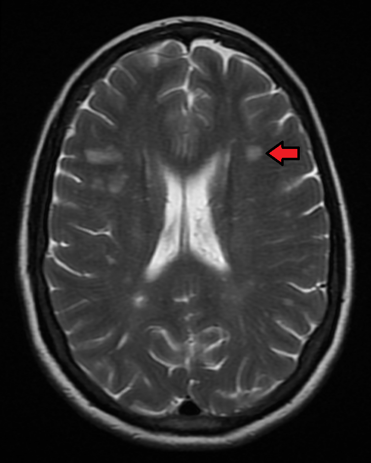
- MRI is the test of choice: T2/FLAIR ovoid periventricular lesions perpendicular to the ventricles = Dawson fingers; also juxtacortical, infratentorial, and spinal-cord plaques
- Gadolinium enhancement marks active lesions; simultaneous enhancing plus non-enhancing lesions on a single scan satisfy DIT
- CSF: CSF-specific oligoclonal bands (IgG present in CSF but not serum) with an elevated IgG index — under 2017 criteria OCBs can substitute for DIT; cells usually fewer than 50 lymphocytes, protein normal or mildly elevated
- Visual evoked potentials: delayed latency with preserved amplitude
- Always exclude mimics: AQP4/MOG antibodies, B12, HIV, RPR, sarcoidosis
Demyelinating Disease Comparison
| Feature | Multiple sclerosis | NMOSD (Devic) | ADEM |
|---|---|---|---|
| Antibody | none specific | AQP4-IgG | none specific (MOG-IgG in a subset) |
| Typical patient | young woman | woman (often non-white) | child, post-infection/vaccine |
| Optic neuritis | unilateral, milder | bilateral, severe | may occur |
| Spinal cord | short-segment | longitudinally extensive (3 or more segments) | may occur |
| Signature clue | Dawson fingers, INO | area postrema — intractable hiccups/vomiting | encephalopathy, monophasic |
| CSF oligoclonal bands | positive (~85%) | usually negative | often negative |
| First-line Rx | disease-modifying therapy | rituximab, eculizumab; avoid interferon-beta | IV steroids |
Charcot's neurologic triad of MS — 'SIN':
- S — Scanning (staccato) speech
- I — Intention tremor
- N — Nystagmus
Reflects brainstem–cerebellar plaque involvement. Do not confuse it with Charcot's cholangitis triad (fever, jaundice, RUQ pain) — same eponym, different organ.
Management
MS treatment has three arms.
1. Acute relapse: high-dose IV methylprednisolone shortens the attack (no effect on long-term disability); plasma exchange (PLEX) rescues severe, steroid-refractory relapses.
2. Disease-modifying therapy (DMT) lowers relapse rate and new lesions:
- Injectables: interferon-beta, glatiramer acetate
- Orals: dimethyl fumarate, teriflunomide (teratogenic, hepatotoxic), and S1P modulators fingolimod/siponimod — watch first-dose bradycardia and macular edema
- High-efficacy monoclonals: natalizumab (anti-alpha4-integrin; screen JC virus for PML risk) and anti-CD20 ocrelizumab/ofatumumab — ocrelizumab is the only DMT approved for primary progressive MS
3. Symptomatic: spasticity to baclofen/tizanidine; urinary urgency to oxybutynin; fatigue to amantadine; neuropathic pain/trigeminal neuralgia to carbamazepine/gabapentin; slow gait to dalfampridine.
Vignette: A 34-year-old woman develops simultaneous bilateral severe optic neuritis and paraparesis. Cord MRI shows a contiguous lesion from T4–T9 (longitudinally extensive transverse myelitis); CSF oligoclonal bands are negative.
- Diagnosis: neuromyelitis optica spectrum disorder (NMOSD / Devic) — not typical MS.
- Next best step: serum AQP4-IgG (aquaporin-4) antibody; send MOG-IgG if negative.
- Management: acute IV steroids plus/minus PLEX; long-term rituximab, eculizumab, satralizumab, or inebilizumab.
- Board trap: do NOT give interferon-beta, natalizumab, or fingolimod — MS DMTs can worsen NMOSD.
Practice Neuro now
Board-style questions, spaced-repetition flashcards, and a Socratic AI tutor — free to start.