Multiple Myeloma & Plasma Cell Disorders
A boards-focused walkthrough of multiple myeloma and related plasma cell disorders — from the 'one clone, one antibody' pathophysiology and CRAB presentation through SPEP/free light chains, marrow criteria, and high-risk cytogenetics, with next-best-step vignettes and a MGUS/smoldering/MM/Waldenström comparison table.
Pathophysiology: one clone, one antibody, one light chain
Multiple myeloma (MM) is a malignant clonal proliferation of terminally differentiated B-cells — plasma cells — in the bone marrow that secretes a single monoclonal immunoglobulin (M protein/paraprotein), most often IgG > IgA. Almost every board finding follows from one idea: one clone makes one antibody and one light chain.
- The clone crowds out marrow → anemia and functional hypogammaglobulinemia (deficient useful polyclonal antibody despite ↑ total Ig) → infection.
- Myeloma cells secrete cytokines — IL-6 (key growth factor), RANKL, MIP-1α — that activate osteoclasts and suppress osteoblasts (via DKK1), producing purely lytic ('punched-out') bone lesions and hypercalcemia.
- Excess free light chains (Bence Jones protein, κ or λ) are filtered, then precipitate in tubules → cast nephropathy ('myeloma kidney').
MM is a disease of older adults (median ~65–70) and is more common in African Americans. It sits on a spectrum: MGUS → smoldering MM → symptomatic MM.
- CRAB = myeloma-defining organ damage: Calcium ↑, Renal failure, Anemia, Bone lesions.
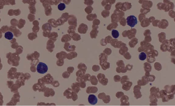
- Anemia is normocytic; smear shows rouleaux (RBCs stacked like coins) from high paraprotein; markedly ↑ ESR.
- ↑ Total serum protein with normal/low albumin = wide globulin gap (the M protein is globulin, not albumin).
- Bone lesions are LYTIC / 'punched-out' — image with whole-body low-dose CT, PET-CT, or MRI (now preferred over the older skeletal survey). Radionuclide bone scan is falsely negative (no osteoblastic reaction).
- Infection is the leading cause of death — encapsulated organisms (S. pneumoniae, H. influenzae).
- M spike on SPEP; urine Bence Jones light chains are missed by dipstick (dipstick detects albumin only — use sulfosalicylic acid or UPEP).
- Complications: AL amyloidosis (light-chain deposition) and renal failure.
CRAB — the four myeloma-defining organ injuries:
- C — hyperCalcemia ('stones, bones, groans, psychiatric overtones')
- R — Renal insufficiency (light-chain cast nephropathy)
- A — Anemia (normocytic; marrow replacement)
- B — Bone lytic lesions (back pain, pathologic fractures)
High-yield board buzzwords to pair with it:
- Bence Jones = urinary free light chains
- Rouleaux = stacked RBCs on smear
- 'Punched-out' = lytic skull lesions on X-ray
A 68-year-old man has 3 months of progressive low back pain and fatigue. Labs: Hb 8.9, Ca 11.8, creatinine 2.4, total protein 9.5 with albumin 3.1. Peripheral smear shows RBCs stacked in rows. Skull X-ray shows multiple round lucencies.
- Most likely diagnosis: Multiple myeloma (CRAB + rouleaux + punched-out skull lesions).
- Best next step (initial workup): SPEP + UPEP with immunofixation and a serum free light chain (FLC) assay to identify and quantify the M protein.
- Confirm with: bone marrow biopsy → ≥10% clonal plasma cells.
- Do NOT order a routine bone scan — lytic lesions are missed; use whole-body low-dose CT or MRI.
- Framing: the CRAB features here (Ca 11.8, Cr 2.4, Hb 8.9, lytic lesions) are myeloma-defining organ damage — this is symptomatic MM, not MGUS or smoldering MM.
Comparison: MGUS vs smoldering MM vs MM vs Waldenström
| Disorder | M protein | Marrow clonal cells | Organ damage | Board clue |
|---|---|---|---|---|
| MGUS | < 3 g/dL | < 10% plasma cells | None | Asymptomatic; ~1%/yr → MM |
| Smoldering MM | ≥ 3 g/dL | 10–60% plasma cells | None | Asymptomatic; ~10%/yr (first 5 yr) |
| Multiple myeloma | Any (usually IgG > IgA) | ≥ 10% (or plasmacytoma) + MDE | CRAB or SLiM | Lytic bone, rouleaux, Bence Jones |
| Waldenström | IgM | Lymphoplasmacytic cells | Hyperviscosity; NO lytic bone | MYD88 L265P; plasmapheresis |
- Diagnostic criteria (IMWG): clonal marrow plasma cells ≥ 10% (or biopsy-proven plasmacytoma) PLUS ≥ 1 myeloma-defining event — CRAB or SLiM.
- SPEP: tall, narrow M spike in the γ region; immunofixation identifies the isotype.
- Serum free light chains (κ/λ ratio): sensitive; detects light-chain-only myeloma.
- Marrow/smear: blood rouleaux; marrow shows sheets of plasma cells — eccentric nucleus, perinuclear hof, 'clock-face' chromatin. Russell bodies (cytoplasmic Ig), Dutcher bodies (nuclear Ig invaginations).
- Staging/prognosis: β2-microglobulin + albumin → ISS (R-ISS adds LDH + high-risk cytogenetics).
- High-risk FISH cytogenetics: del(17p), t(4;14), t(14;16), gain(1q); hyperdiploidy and t(11;14) = standard/favorable risk.
- Pitfall: correct total calcium for low albumin (or check ionized Ca).

A 70-year-old woman has headache, blurred vision, epistaxis, and dizziness. Fundoscopy shows dilated, tortuous 'sausage-link' retinal veins. Total protein is high; SPEP reveals an IgM M spike. There are no lytic bone lesions and no hypercalcemia.
- Diagnosis: Waldenström macroglobulinemia (lymphoplasmacytic lymphoma) with hyperviscosity syndrome from large pentameric IgM.
- Mutation: MYD88 L265P.
- Best next step (symptomatic hyperviscosity): plasmapheresis (plasma exchange) to rapidly remove IgM — before starting rituximab-based therapy (rituximab can trigger an IgM flare).
- Key contrast with MM: IgM (not IgG/IgA), hyperviscosity + NO lytic bone. MM rarely causes hyperviscosity because IgG/IgA are smaller and less viscous than pentameric IgM.
Management principles (know the mechanisms)
Treat the clone and protect end organs. Modern first-line regimens combine a proteasome inhibitor (bortezomib) + an immunomodulatory drug (lenalidomide/thalidomide) + dexamethasone (VRd); transplant-eligible patients proceed to autologous stem cell transplant.
- Bortezomib blocks the proteasome — plasma cells synthesize enormous amounts of Ig and depend on proteasomal clearance of misfolded protein, so they are exquisitely sensitive.
- Thalidomide/lenalidomide are antiangiogenic/immunomodulatory — teratogens (thalidomide → phocomelia).
- Bone disease: bisphosphonates (or denosumab) reduce fractures/pain — watch for osteonecrosis of the jaw.
- Acute care: aggressive IV hydration for hypercalcemia and to protect kidneys; avoid nephrotoxins/IV contrast.
MM is incurable but highly treatable; MGUS is only observed (no treatment).
Practice Heme/Onc now
Board-style questions, spaced-repetition flashcards, and a Socratic AI tutor — free to start.