Microcytic Anemias
A board-focused walk through the microcytic anemias — iron deficiency, anemia of chronic disease, thalassemia, and sideroblastic/lead — anchored on the classic iron-studies pattern, peripheral-smear clues, and the TAILS framework.
What Makes a Cell Microcytic
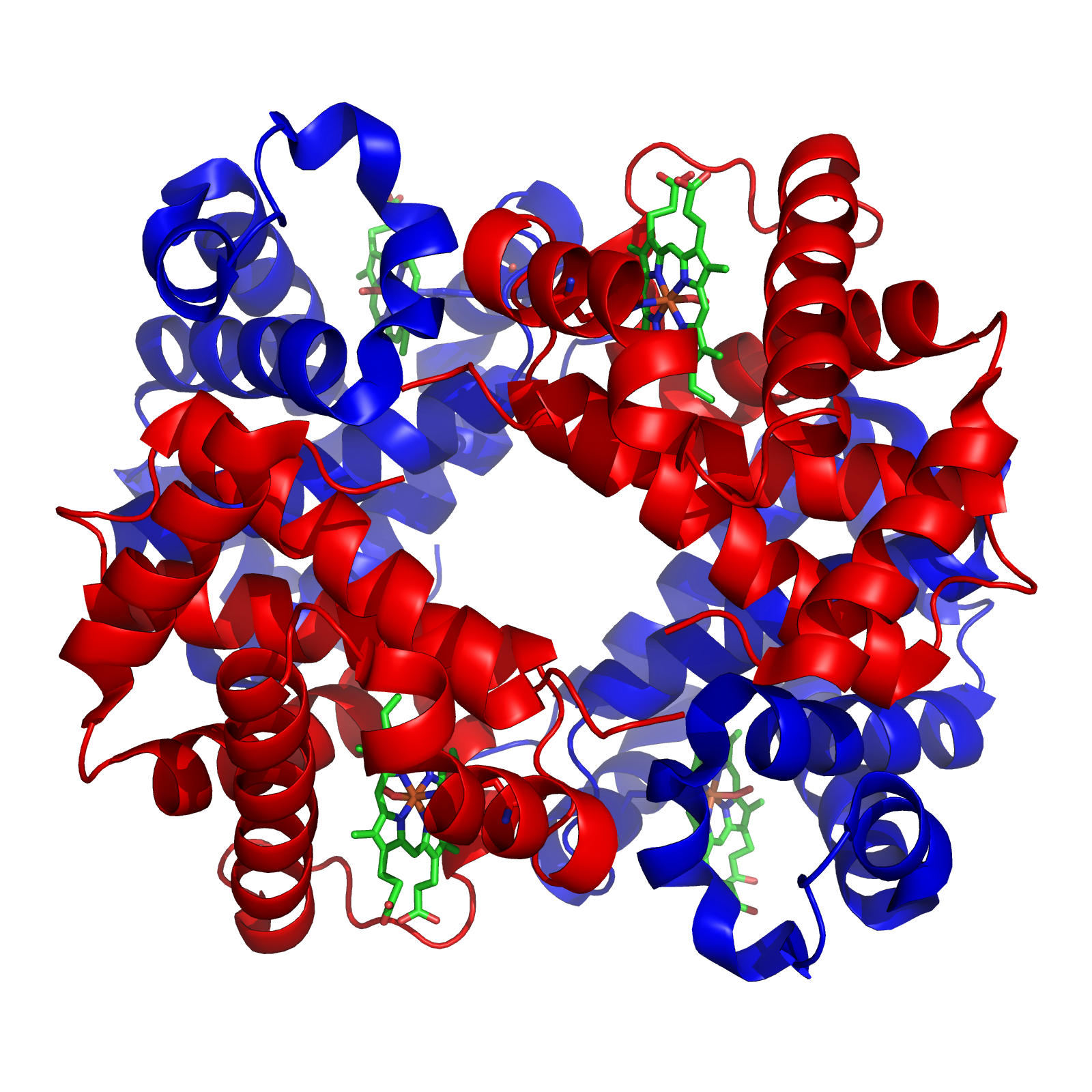
Microcytic anemia is defined by a mean corpuscular volume (MCV) < 80 fL. The final act of erythropoiesis is packing each red cell with hemoglobin; when a building block runs short, precursors keep dividing to reach a threshold hemoglobin concentration, yielding small (microcytic), pale (hypochromic) cells. Hemoglobin has two parts — heme (iron + a protoporphyrin ring) and four globin chains — so every microcytic anemia is a failure of one of three things: iron supply (iron-deficiency anemia, anemia of chronic disease), protoporphyrin/heme synthesis (sideroblastic anemia, lead poisoning), or globin synthesis (the thalassemias).
- Iron-deficiency anemia (IDA) is the most common cause of microcytic anemia and the most common anemia worldwide.
- Ferritin is the single most useful test: a low ferritin is essentially diagnostic of iron deficiency. Because ferritin is an acute-phase reactant (and iron stores are genuinely replete in the other microcytic anemias), it runs normal-to-high in ACD, thalassemia, and sideroblastic anemia — so a low ferritin rules IDA *in*, but a normal/high ferritin does NOT rule it *out* when inflammation coexists.
- New IDA in an adult male or postmenopausal female = occult GI blood loss (colon cancer) until proven otherwise → colonoscopy.
- Thalassemia clue: normal-to-high RBC count with a disproportionately low MCV and a normal RDW. Mentzer index (MCV ÷ RBC) < 13 favors thalassemia; > 13 favors IDA.
- ↑ HbA2 (> 3.5%) on electrophoresis confirms β-thalassemia minor.
- Basophilic stippling → think lead poisoning, sideroblastic anemia, or thalassemia.
- Ringed sideroblasts (iron-laden mitochondria ringing the nucleus, Prussian-blue stain) define sideroblastic anemia.
Iron Studies at a Glance
| Study | Iron deficiency | Chronic disease | Thalassemia | Sideroblastic |
|---|---|---|---|---|
| Serum iron | ↓ | ↓ | normal | ↑ |
| TIBC (transferrin) | ↑ | ↓ | normal | ↓ / normal |
| Ferritin | ↓ | ↑ | normal | ↑ |
| Transferrin saturation | ↓ | ↓ / normal | normal | ↑ |
| RDW | ↑ | normal | normal | ↑ |
Classic vignette: a menstruating woman, a toddler on excessive cow's milk, or an older adult with fatigue, pallor, and exertional dyspnea. The buzzwords:
- Pica — craving ice (pagophagia), dirt, or starch
- Koilonychia (spoon-shaped nails), angular cheilosis, atrophic glossitis
- Plummer-Vinson syndrome = IDA + esophageal webs + atrophic glossitis → dysphagia (also ↑ esophageal SCC risk)
- Smear: hypochromic microcytic cells with pencil (cigar) cells and marked anisocytosis (↑ RDW)
Next best step / management:
- Confirm with iron studies (↓ ferritin is the earliest, most specific marker).
- Find the source — in adult men and postmenopausal women, pursue GI evaluation (colonoscopy ± EGD) for occult malignancy.
- Treat with oral ferrous sulfate; a reticulocytosis within ~1 week confirms response, with hemoglobin and MCV normalizing over weeks to months.

The Thalassemias
Thalassemias are inherited defects in the rate of globin-chain synthesis; the resulting chain imbalance precipitates and damages RBCs (ineffective erythropoiesis + hemolysis). α-thalassemia arises from deletions of α-globin genes (4 total, chromosome 16), and severity tracks the number lost. β-thalassemia arises from point mutations (promoter/splice-site) in the β-globin gene (chromosome 11).
Because β-chains are needed for adult HbA (α2β2), β-thalassemia major only becomes symptomatic around 6 months of age, as protective fetal HbF (α2γ2) declines. Chronic ineffective erythropoiesis expands the marrow — 'crew-cut' skull on X-ray and chipmunk facies — and drives extramedullary hematopoiesis with hepatosplenomegaly. Lifelong transfusions then cause secondary iron overload (hemochromatosis), managed with iron chelation. Smear shows target cells. One α-thal genetics pearl: Asian carriers tend to have both deletions on the same chromosome (cis), risking severe (HbH / Hb Barts) offspring, whereas African carriers usually carry them in trans.
Thalassemia: Genotype → Phenotype
| Type | Genetic defect | Clinical picture |
|---|---|---|
| α-thal silent carrier | 1 of 4 α-genes deleted | Asymptomatic, normal labs |
| α-thal trait (minor) | 2 α-genes deleted | Mild microcytic anemia, no treatment |
| HbH disease | 3 α-genes deleted → HbH (β4) | Moderate–severe hemolytic anemia |
| Hb Barts | 4 α-genes deleted → Hb Barts (γ4) | Hydrops fetalis, fatal in utero |
| β-thal minor (trait) | Heterozygous β point mutation | Mild anemia, ↑ HbA2 (> 3.5%) |
| β-thal major (Cooley) | Homozygous β⁰ | Severe, transfusion-dependent, ↑ HbF & HbA2 |

Sideroblastic Anemia & Lead Poisoning
Sideroblastic anemia is defective heme synthesis: iron enters erythroblast mitochondria but can't be incorporated into heme (protoporphyrin synthesis is impaired), so it accumulates as ringed sideroblasts. Iron studies therefore mimic overload (↑ iron, ↑ ferritin, ↑ transferrin saturation). Causes are hereditary (X-linked δ-ALA synthase / ALAS2 defect) or acquired — reversible causes include alcohol (most common overall), lead, isoniazid (depletes vitamin B6 / pyridoxine, the ALA-synthase cofactor), and copper deficiency; the key clonal (irreversible) cause is myelodysplastic syndrome. Pyridoxine-responsive (often hereditary) forms improve with B6.
Lead poisoning inhibits ferrochelatase and ALA dehydratase, blocking heme synthesis (effectively an acquired sideroblastic anemia). Boards clues: a child with pica in an old building (paint/dust) with abdominal pain, constipation, and encephalopathy, plus basophilic stippling on smear; adults get peripheral neuropathy (wrist/foot drop) and gingival Burton (lead) lines. Treat with succimer (oral, children) or EDTA + dimercaprol (BAL) for severe or symptomatic poisoning.
Causes of microcytic anemia — "TAILS":
- T — Thalassemia
- A — Anemia of chronic disease (classically normocytic; becomes microcytic when chronic)
- I — Iron deficiency
- L — Lead poisoning
- S — Sideroblastic anemia
Practice Heme/Onc now
Board-style questions, spaced-repetition flashcards, and a Socratic AI tutor — free to start.