The Mediastinum & Thoracic Anatomy
A board-focused tour of the mediastinum: the sternal-angle master landmark, the four divisions and their contents, and the high-yield clinical correlations — left recurrent laryngeal palsy, SVC syndrome, chylothorax, and the phrenic-anterior/vagus-posterior lung-root rule. Built for USMLE Step 1 vignettes.
The mediastinum & the sternal angle of Louis
The mediastinum is the median partition of the thorax between the two pleural sacs, running from the thoracic inlet to the diaphragm and from the sternum to the vertebral column. The sternal angle (of Louis) — the palpable transverse ridge at the manubriosternal joint, opposite the 2nd costal cartilage and the T4/T5 disc — is the single most tested landmark. The horizontal plane through it splits the superior mediastinum (above) from the inferior mediastinum (below), which the pericardial sac further divides into anterior, middle, and posterior compartments. At this one plane the arch of the aorta begins and ends, the trachea bifurcates (carina), the azygos vein arches over the right main bronchus into the SVC, the ligamentum arteriosum and cardiac plexus sit, and the thoracic duct crosses from right to left.
The four divisions and what lives in each
| Division | Boundaries | Key contents | What the boards test |
|---|---|---|---|
| Superior | thoracic inlet → sternal-angle plane (T4/T5) | arch of aorta + its 3 branches, brachiocephalic veins, upper SVC, trachea, esophagus, thoracic duct, thymic remnant, vagus, phrenic, left recurrent laryngeal nn. | left RLN palsy → hoarseness; thymoma |
| Anterior | sternum → fibrous pericardium | thymus, fat, lymph nodes, internal thoracic vessels | anterior mass = the 4 T's |
| Middle | within the pericardial sac | heart + pericardium, roots of great vessels, SVC, main bronchi, phrenic nn., pulmonary vessels | cardiac tamponade; SVC syndrome |
| Posterior | pericardium → T5–T12 vertebrae | descending thoracic aorta, esophagus, thoracic duct, azygos/hemiazygos, vagus, sympathetic trunks & splanchnic nn. | posterior mass = neurogenic tumor; aortic aneurysm |
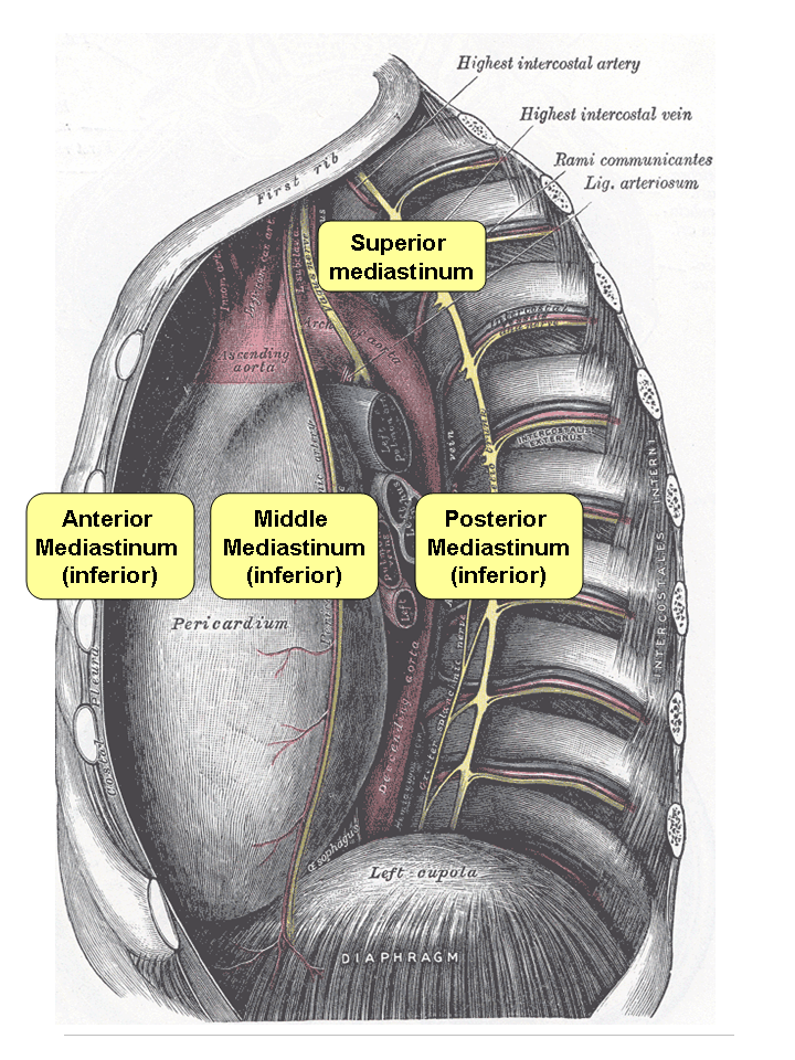
- Sternal angle = T4/T5: aortic arch start/end, tracheal bifurcation (carina), azygos arch, ligamentum arteriosum, superior↔inferior mediastinum boundary.
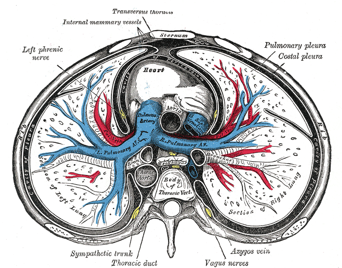
- Phrenic nerve (C3–C5) runs ANTERIOR to the lung root (on the fibrous pericardium); the vagus runs POSTERIOR to the lung root — a favorite exam discriminator.
- Aortic arch branches, proximal → distal: brachiocephalic trunk → left common carotid → left subclavian.
- Diaphragm apertures: IVC (+ right phrenic n.) at T8; esophagus (+ vagal trunks) at T10; aorta (+ thoracic duct + azygos vein) at T12.
- Azygos vein arches over the right lung root; the aortic arch arches over the left lung root.
- Diaphragm apertures — "I 8, 10 EGGs, AAT 12": vena cava at T8, Esophagus + vaGus at T10, Aorta + Azygos + Thoracic duct at T12.
- Phrenic roots — "C3, 4, 5 keep the diaphragm alive."
- Anterior mediastinal mass — the 4 T's: Thymoma, Teratoma (germ-cell), Thyroid (retrosternal goiter), Terrible lymphoma.
- Lung-root artery position — "RALS": the pulmonary artery is Right-Anterior and Left-Superior relative to the main bronchus.
A 62-year-old smoker has months of progressive hoarseness; CT shows a left hilar mass. The left recurrent laryngeal nerve leaves the vagus, hooks under the arch of the aorta just lateral to the ligamentum arteriosum, then ascends in the tracheo-esophageal groove to the larynx. Its long intrathoracic loop makes it vulnerable to left hilar bronchogenic carcinoma, aortic-arch aneurysm, and left atrial enlargement from mitral stenosis (Ortner cardiovocal syndrome). Injury paralyzes all intrinsic laryngeal muscles except the cricothyroid (external branch of the superior laryngeal n.) → a hoarse, breathy voice with a paramedian vocal cord. (The right RLN loops under the right subclavian artery and is spared by arch pathology — suspect thyroid/neck surgery or an apical lung tumor instead.)
A patient with small-cell lung cancer develops facial plethora, periorbital and upper-limb edema, distended non-pulsatile neck veins, and a headache worse on bending forward. The superior vena cava is thin-walled and low-pressure, so a right-sided mediastinal mass readily compresses it — most often bronchogenic carcinoma (especially small-cell), lymphoma, or thymoma. Venous return detours through the azygos system, the key SVC–IVC collateral. This is SVC syndrome: an oncologic urgency and a classic boards vignette, usually driven by extrinsic compression rather than an immediate airway emergency.
Nerves of the mediastinum → course → injury
| Nerve | Root / origin | Course & key relation | Function | Injury → deficit |
|---|---|---|---|---|
| Phrenic | C3–C5 (mainly C4) | anterior to lung root, on fibrous pericardium | sole motor to diaphragm; sensory to pericardium + mediastinal/diaphragmatic pleura | paralyzed hemidiaphragm (elevated, paradoxical rise on sniff); referred shoulder-tip pain |
| Vagus (CN X) | medulla | posterior to lung root; forms esophageal plexus → anterior (L) & posterior (R) vagal trunks | parasympathetic to thoracic & foregut viscera | — (named branches matter more) |
| Left recurrent laryngeal | vagus | hooks under aortic arch, lateral to lig. arteriosum; ascends in T-E groove | intrinsic laryngeal muscles (except cricothyroid) | hoarseness — arch aneurysm, left hilar Ca, mitral stenosis (Ortner) |
| Right recurrent laryngeal | vagus | hooks under right subclavian artery | same | hoarseness (thyroid/neck surgery, apical tumor) |
| Sympathetic trunk / splanchnics | T1–T12 paravertebral chain (greater T5–9, lesser T10–11, least T12) | trunk on the rib heads (lateral to the vertebral bodies); splanchnics cross the bodies to pierce the crura | sympathetic to thoracic & abdominal viscera; T1 fibres to head/eye | apical Pancoast tumor invading the chain → Horner (ptosis, miosis, anhidrosis) |
- Three anatomical constrictions (where foreign bodies lodge and caustic/reflux strictures form), measured from the incisors: (1) cricopharyngeus / upper esophageal sphincter (~C6, ~15 cm); (2) crossing of the aortic arch + left main bronchus (~22–27 cm); (3) esophageal hiatus / LES (T10, ~40 cm).
- Pierces the diaphragm at T10 with the anterior (left) and posterior (right) vagal trunks.
- Retrocardiac position: left atrial enlargement (mitral stenosis) can indent the esophagus on barium swallow.
- The lower third drains to the left gastric → portal vein — the portosystemic anastomotic site for esophageal varices.
During esophageal or thoracic surgery (or penetrating trauma), the thoracic duct is torn → chylothorax: a milky pleural effusion rich in triglycerides and chylomicrons. The duct arises from the cisterna chyli (~L1/L2), enters the thorax through the aortic hiatus (T12), ascends on the right of the vertebral bodies, then crosses to the left at ~T4/T5 to empty into the left venous angle (junction of the left subclavian and internal jugular veins). Because of this crossover, a lower duct injury tends to produce a right chylothorax and an upper injury a left one. The duct drains lymph from the whole body except the right upper quadrant, which drains via the right lymphatic duct.

Big picture: pericardial sinuses & the lung root
Two pericardial recesses in the middle mediastinum are testable. The transverse pericardial sinus lies behind the ascending aorta and pulmonary trunk and in front of the SVC and atria — a surgeon passes a finger or ligature through it to clamp the arterial outflow during cardiac surgery. The oblique sinus is a blind cul-de-sac behind the left atrium. At the lung root, remember the relations that unlock most vignettes: the phrenic nerve and pericardiacophrenic vessels pass anteriorly, the vagus passes posteriorly, the azygos vein arches over the right root, and the aortic arch over the left. Anchor everything to the sternal-angle plane and these root relations, and mediastinal questions resolve quickly.
Practice Anatomy now
Board-style questions, spaced-repetition flashcards, and a Socratic AI tutor — free to start.