Management of Hyperkalemia
A management-focused Step 2 CK lesson on acute hyperkalemia: the ECG-driven emergency approach and the stabilize–shift–remove algorithm, with confident doses, a decision table, two \"next best step\" vignettes, a mnemonic, and high-yield pitfalls.
Overview & Immediate Approach
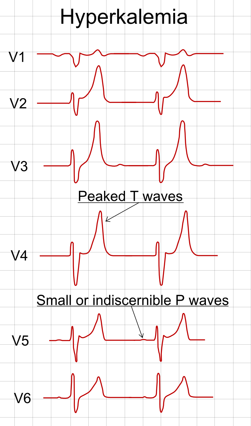
Hyperkalemia (serum K⁺ > 5.5 mEq/L; life-threatening when > 6.5 mEq/L or with any ECG changes) is one of the few electrolyte emergencies where the next best step is driven by the ECG, not the number. The first three moves in any suspected case are: (1) obtain a 12-lead ECG, (2) place the patient on a cardiac monitor, and (3) stop all exogenous K⁺ and K⁺-retaining drugs (ACEi/ARB, spironolactone/K⁺-sparing diuretics, NSAIDs, trimethoprim, K⁺ supplements).
Management then follows three sequential goals — stabilize the myocardium → shift K⁺ into cells → remove K⁺ from the body. This order matters because the shifting agents only buy time (minutes to hours); they do not eliminate potassium, so every patient ultimately needs a removal strategy. If the value could be spurious (hemolysis, prolonged tourniquet/fist-clenching, marked leukocytosis/thrombocytosis) and the patient is asymptomatic with a normal ECG, confirm with a repeat draw before treating.

- ECG changes = emergency. Progression: peaked T waves → PR/QRS widening → loss of P waves → sine wave → VF/asystole. Treat the ECG; don't wait for a repeat K⁺.
- IV calcium is the FIRST drug when ECG changes are present — it stabilizes the cardiac membrane within minutes but does NOT lower serum K⁺.
- Insulin + glucose is the mainstay for shifting K⁺ intracellularly; add high-dose nebulized albuterol for an additive effect.
- Only insulin, albuterol, and (in coexisting metabolic acidosis) bicarbonate SHIFT K⁺; loop diuretics, GI binders, and dialysis REMOVE it — every patient eventually needs removal.
- Hemodialysis is definitive — the priority in ESRD, oliguric AKI, or refractory/severe hyperkalemia.
- Recheck K⁺ and glucose after treatment — insulin-induced hypoglycemia is common and can occur hours later.
Stepwise Management Algorithm
| Priority | Intervention | Typical dose | Onset | Effect on serum K⁺ |
|---|---|---|---|---|
| 1. Stabilize membrane | IV calcium gluconate 10% | 10 mL (1 g) IV over 2–3 min; repeat in 5 min if ECG unchanged | 1–3 min | None — cardioprotective only |
| 2. Shift into cells | Regular insulin + dextrose | 10 units IV insulin + 25 g dextrose (D50); monitor glucose | 10–20 min | ↓ ~0.5–1 mEq/L for 4–6 h |
| Nebulized albuterol | 10–20 mg neb (much higher than the asthma dose) | ~30 min | ↓ ~0.5–1 mEq/L; additive to insulin | |
| Sodium bicarbonate | Only if metabolic acidosis present | slow/variable | Adjunct; unreliable alone | |
| 3. Remove from body | Loop diuretic (furosemide) | if urine output adequate | 15–60 min | ↑ urinary K⁺ excretion |
| GI binder: Na⁺ zirconium cyclosilicate or patiromer | non-emergent dosing | hours | Subacute/chronic lowering | |
| Hemodialysis | — | minutes | Definitive for refractory/ESRD |
A 58-year-old man with ESRD on hemodialysis misses two sessions and presents with generalized weakness. K⁺ is 7.4 mEq/L; ECG shows peaked T waves with a widened QRS.
Next best step: IV calcium gluconate (10 mL of 10% over 2–3 min) immediately to stabilize the myocardium — before insulin, before dialysis. Repeat in 5 min if the ECG has not improved.
Then: insulin 10 units IV + 25 g dextrose and nebulized albuterol to shift K⁺, and arrange emergent hemodialysis (definitive therapy given anuric ESRD). Loop diuretics and GI binders are useless here — he makes little to no urine, and binders act over hours.
"C BIG K" — the agents of hyperkalemia therapy:
- C — Calcium (membrane stabilization; first if ECG changes)
- B — Beta-agonist (albuterol) and Bicarbonate (only if acidotic)
- I — Insulin
- G — Glucose (always with insulin unless already hyperglycemic)
- K — K⁺ removal: GI binders (Kayexalate is slow/risky — prefer Na⁺ zirconium cyclosilicate or patiromer), loop diuretics (kidneys), and dialysis (definitive)
Memory hook: the first four (C-B-I-G) stabilize and shift; only the final K actually removes potassium from the body — so a patient given C-B-I-G alone is not yet definitively treated.
A 44-year-old asymptomatic woman has a routine K⁺ of 6.3 mEq/L. The lab flags the specimen as hemolyzed. She takes no medications, renal function is normal, and her ECG is completely normal.
Next best step: Repeat the potassium with a properly collected, non-hemolyzed specimen before any treatment. Hemolysis, prolonged tourniquet/fist-clenching, and marked thrombocytosis or leukocytosis cause pseudohyperkalemia. Treating an unconfirmed spurious value risks iatrogenic hypokalemia.
Contrast: if ECG changes were present, you would treat first (calcium + shifting) and confirm the level in parallel — never delay therapy in a symptomatic or ECG-positive patient.
- Calcium does not lower K⁺ — it only buys time; you must still shift and remove.
- Never give insulin without glucose (unless glucose > 250 mg/dL) — monitor glucose for 4–6 h; delayed hypoglycemia is common.
- Bicarbonate is not effective monotherapy and helps only with a coexisting metabolic acidosis.
- Sodium polystyrene sulfonate (Kayexalate) is slow and can cause colonic necrosis (especially with sorbitol or postoperatively) — avoid for acute emergencies; newer binders (Na⁺ zirconium cyclosilicate, patiromer) are preferred for subacute/chronic control.
- Always remove the cause: hold ACEi/ARB, K⁺-sparing diuretics, NSAIDs, trimethoprim, non-selective β-blockers, and K⁺ supplements.
- Escalate to dialysis early in ESRD, oliguric AKI, or when K⁺ remains high despite medical therapy.
Practice Clinical Management now
Board-style questions, spaced-repetition flashcards, and a Socratic AI tutor — free to start.