Management of Acute GI Bleeding
A Step 2 CK-focused walkthrough of acute GI bleeding management: resuscitate before you investigate, risk-stratify, give the right pre-endoscopy pharmacology (IV PPI for ulcers; octreotide + ceftriaxone for varices), transfuse restrictively, and know exactly when to escalate to endoscopy, IR, or surgery.
The core principle: resuscitate before you investigate
Acute GI bleeding is a hemodynamic emergency, and the exam-tested principle is that stabilization always precedes diagnosis. Whether the source is upper (proximal to the ligament of Treitz) or lower (distal to it), the first moves are identical: ABCs, two large-bore IVs (16–18 gauge), crystalloid resuscitation, and type & crossmatch.
Localizing clues: Hematemesis or coffee-ground emesis = upper source. Melena usually = upper (but can be small-bowel/right colon). Hematochezia usually = lower, but a brisk upper GI bleed can present as hematochezia — so in an unstable patient with red blood per rectum, still consider (and often first exclude) an upper source. A BUN:creatinine ratio > 20–30 points toward an upper source (digested blood is an absorbed nitrogen load).
On a "next best step" question in an unstable bleeder, the answer is almost always resuscitation (IV access, fluids, blood) or airway protection — not "order endoscopy" or "order CT."
- Two large-bore IVs (16–18 g), crystalloid resuscitation, type & cross; send CBC, BMP, LFTs, INR/coags, lactate
- Restrictive transfusion: transfuse PRBCs for Hgb < 7 g/dL (target 7–9); use a higher < 8 threshold in cardiovascular disease/ACS. In massive hemorrhage with shock, give blood based on ongoing losses — don't wait on an early, falsely-normal Hgb. Over-transfusing stable UGIB raises rebleeding/mortality
- Suspected peptic-ulcer (non-variceal) UGIB → empiric IV PPI (e.g., pantoprazole)
- Suspected variceal bleed (cirrhosis) → octreotide + prophylactic IV ceftriaxone before endoscopy — antibiotics reduce mortality
- EGD within 24 h for UGIB (within ~12 h for suspected variceal); colonoscopy for stable LGIB after prep
- Erythromycin IV before EGD acts as a prokinetic to clear the stomach and improve visualization
- Reverse coagulopathy (hold/reverse anticoagulants; 4-factor PCC + IV vitamin K for warfarin; platelets if < 50k and bleeding) — but never delay resuscitation to do so
- Glasgow-Blatchford score 0–1 → very low risk; consider outpatient management
- Massive/unstable LGIB → first exclude an upper source (EGD), then CT angiography → angiographic embolization; surgery if refractory (colonoscopy is for stable patients after prep)
- Routine NG lavage is no longer required and does not change outcomes
The ordered algorithm (upper GI bleed)
- Resuscitate: ABCs, two large-bore IVs, crystalloid, type & cross. Secure the airway (intubate) if the patient is obtunded or has massive ongoing hematemesis.
- Labs & risk-stratify: CBC, BMP (↑BUN:Cr suggests upper source), LFTs, INR/coags; calculate the Glasgow-Blatchford score (0–1 = low risk, potential outpatient).
- Transfuse restrictively: PRBC for Hgb < 7 (target 7–9); higher (< 8) threshold in CAD/ACS. If in hemorrhagic shock, give blood immediately regardless of the initial Hgb. Correct coagulopathy/thrombocytopenia.
- Pharmacology: IV PPI for suspected ulcer; if cirrhosis/varices suspected add octreotide + ceftriaxone; consider erythromycin pre-scope.
- Endoscopy within 24 h (~12 h for variceal) — both diagnostic and therapeutic.
- Escalate if uncontrolled: repeat EGD → angiographic embolization (IR) for non-variceal; TIPS for refractory variceal bleeding; surgery as last resort.
Variceal vs. non-variceal (peptic ulcer) UGIB
| Feature | Variceal bleed | Peptic ulcer (non-variceal) |
|---|---|---|
| Typical setting | Cirrhosis / portal hypertension | NSAIDs, H. pylori |
| Pre-endoscopy drug | Octreotide 50 mcg IV bolus → 50 mcg/h | IV PPI (pantoprazole) |
| Antibiotics | Yes — ceftriaxone (↓ mortality, ↓ SBP/infection) | No |
| Endoscopic therapy | Band ligation (first line) | Dual therapy: epinephrine injection + clip or thermal (never epinephrine alone) |
| Escalation / rescue | Balloon tamponade (bridge) → TIPS | Repeat EGD → angioembolization → surgery |
| Secondary prevention | Nonselective beta-blocker + serial band ligation | High-dose PPI, treat H. pylori, stop NSAIDs |
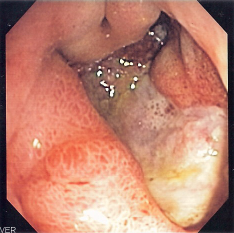
A 55-year-old man with alcoholic cirrhosis presents with two episodes of large-volume hematemesis. BP 86/52, HR 122; he is pale and diaphoretic.
Immediate steps: two large-bore IVs, crystalloid, type & cross, transfuse toward Hgb 7–9 (give blood now given shock, don't wait on the first Hgb).
Next best step in management: Start IV octreotide (splanchnic vasoconstriction) and IV ceftriaxone (prophylactic antibiotics reduce mortality and infection in cirrhotic bleeders), then proceed to urgent EGD within 12 hours for endoscopic variceal band ligation. If bleeding is uncontrolled, use balloon tamponade as a bridge and arrange TIPS.
Trap: Antibiotics are the most commonly forgotten, mortality-reducing intervention in variceal bleeding — don't skip them.

AIMS65 is a validated bedside score predicting in-hospital mortality in acute upper GI bleeding (1 point each):
- A — Albumin < 3.0 g/dL
- I — INR > 1.5
- M — altered Mental status
- S — Systolic BP ≤ 90 mmHg
- 65 — age ≥ 65 years
More points → higher mortality; useful for triage (ICU vs. floor). *(Contrast with the Glasgow-Blatchford score, which predicts need for intervention/transfusion and identifies low-risk patients potentially safe for outpatient care.)*
Key drugs and doses
| Agent | When to use | Dose | Notes |
|---|---|---|---|
| Pantoprazole (PPI) | Suspected/confirmed peptic-ulcer UGIB | 80 mg IV bolus, then 8 mg/h infusion (or 40 mg IV BID) | Continue high-dose ×72 h for high-risk ulcer stigmata (active bleeding, visible vessel) |
| Octreotide | Variceal bleed | 50 mcg IV bolus, then 50 mcg/h ×3–5 d | Splanchnic vasoconstriction, lowers portal pressure |
| Ceftriaxone | Any cirrhotic with GI bleed | 1 g IV q24h, up to 7 d | ↓ mortality, ↓ SBP/bacterial infection |
| Erythromycin | ~30–120 min before EGD | 250 mg IV | Prokinetic — clears gastric blood for visualization |
| PRBC transfusion | Hgb < 7 (target 7–9); < 8 if CAD/ACS | Target 7–9 g/dL | Restrictive strategy improves survival; in shock, transfuse by hemodynamics |
A 60-year-old man with known peptic ulcer disease arrives with massive ongoing hematemesis. He is now somnolent, cannot protect his airway, and his SpO₂ is falling as he continues to vomit blood.
Next best step in management: Endotracheal intubation to protect the airway before endoscopy — aspiration of blood is a major cause of morbidity, and a secured airway allows safer EGD. Resuscitation and IV PPI continue in parallel; urgent EGD follows once the airway is secured.
Principle: Airway before scope. In an unstable bleeder, the next step is nearly always resuscitation or airway control — not immediately ordering endoscopy.
- Delaying resuscitation to obtain endoscopy or imaging — stabilize first
- Over-transfusing — a liberal strategy raises mortality/rebleeding in stable UGIB; target Hgb 7–9 (threshold ~8 with CAD/ACS). Conversely, in overt hemorrhagic shock, don't withhold blood waiting on a lagging Hgb
- Assuming melena = active bleeding — melena can persist for days after bleeding has stopped
- Forgetting that a brisk upper GI source can present as hematochezia — in the unstable patient with red blood per rectum, exclude an upper source (EGD) first
- Skipping prophylactic antibiotics in a cirrhotic bleeder (proven mortality benefit)
- Using epinephrine injection as monotherapy — it must be combined with a second modality (clip or thermal)
- Not treating H. pylori / stopping NSAIDs after a peptic-ulcer bleed (secondary prevention)
Practice Clinical Management now
Board-style questions, spaced-repetition flashcards, and a Socratic AI tutor — free to start.