Lymphoid Organs & Immune Cells
A Step 1 high-yield tour of primary vs. secondary lymphoid organ architecture (lymph node, spleen, thymus) and immune cell/molecule functions, mapped to the classic defect-to-disease correlations (DiGeorge, asplenia, CGD, LAD, hyper-IgM, APECED). Includes two comparison tables, two clinical vignettes, and board-standard mnemonics.
How the Immune System Is Organized
Primary vs. secondary lymphoid organs is the framework for this entire topic.
- Primary (central) lymphoid organs — the bone marrow and thymus — are where lymphocytes are generated and mature. B cells complete maturation in the bone marrow; T-cell precursors migrate to the thymus for positive and negative selection.
- Secondary (peripheral) lymphoid organs — lymph nodes, spleen, and MALT (tonsils, Peyer patches, appendix) — are where mature, naïve lymphocytes encounter antigen and launch adaptive responses.
The boards reward you for knowing three things about every compartment: (1) which cell lives there, (2) what selects or activates it there, and (3) which infection or autoimmune pattern appears when that compartment fails.
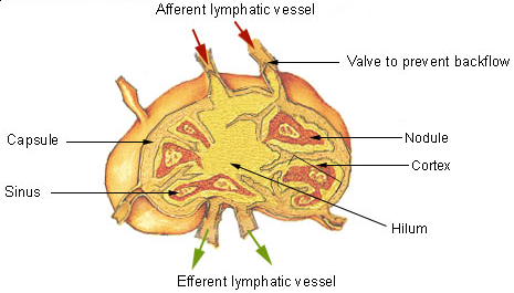
- Lymph node follicle (outer cortex) = B cells. Primary follicles are dense/dormant; secondary follicles have pale germinal centers = active B-cell proliferation and somatic hypermutation.
- Paracortex (between cortex and medulla) = T cells + high endothelial venules (HEV), where circulating lymphocytes enter. Undergoes hyperplasia in viral (cellular) responses; is poorly developed in DiGeorge syndrome and SCID.
- Medulla = medullary cords (plasma cells, B cells) + medullary sinuses (macrophages, reticular cells) draining to efferent lymphatics.
- Spleen white pulp: T cells sit in the periarteriolar lymphatic sheath (PALS); B cells in follicles; the marginal zone holds APCs/macrophages that trap bloodborne antigen.
- Spleen red pulp macrophages remove senescent RBCs and opsonized encapsulated bacteria. The spleen is a major source of IgM and of opsonins (tuftsin, properdin); splenic IgM drives complement activation → C3b opsonization (this is why asplenics can't clear encapsulated organisms).
Lymphoid Compartments — Region → Cell → Clue
| Organ | Region | Predominant cells | Board clue |
|---|---|---|---|
| Lymph node | Follicle (cortex) | B cells | Germinal center = active B-cell proliferation |
| Lymph node | Paracortex | T cells + HEV | Underdeveloped in DiGeorge/SCID; hyperplasia in viral infection |
| Lymph node | Medulla | Plasma cells (cords) + macrophages (sinuses) | Efferent lymph drainage |
| Spleen | White pulp – PALS | T cells | — |
| Spleen | White pulp – follicle / marginal zone | B cells / APCs | Captures bloodborne antigen |
| Spleen | Red pulp | Macrophages | Clears encapsulated bacteria & old RBCs |
| Thymus | Cortex (dense) | Immature thymocytes | Positive selection |
| Thymus | Medulla (pale) | Mature T cells + Hassall corpuscles | Negative selection (AIRE) |
The Thymus & Central Tolerance
The thymus derives from the epithelium of the 3rd pharyngeal pouch (endoderm); its lymphocytes are of mesenchymal (hematopoietic) origin. The cortex is dense with immature thymocytes; the medulla is pale, contains mature T cells and Hassall corpuscles.
- Positive selection (cortex): thymocytes whose TCRs can bind self-MHC survive; those that cannot die by neglect. This ensures MHC restriction.
- Negative selection (medulla): thymocytes whose TCRs bind self-antigen with high affinity undergo apoptosis (clonal deletion) or divert to regulatory T cells — this enforces self-tolerance.
AIRE (autoimmune regulator) drives medullary display of tissue-restricted self-antigens. AIRE mutation → APECED / APS-1: chronic mucocutaneous candidiasis, hypoparathyroidism, and adrenal insufficiency.
- MHC class I (HLA-A, -B, -C): on all nucleated cells (+ platelets, not RBCs); presents endogenous (cytosolic — viral/tumor) peptides loaded via TAP; paired with β2-microglobulin; recognized by CD8⁺ cytotoxic T cells.
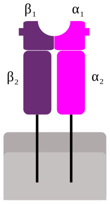
- MHC class II (HLA-DP, -DQ, -DR): on professional APCs (dendritic cells, macrophages, B cells); presents exogenous (phagocytosed) antigen after invariant chain/CLIP exchange in the endosome (HLA-DM–catalyzed); recognized by CD4⁺ helper T cells.
- Rule of 8: MHC I × CD8 = 8; MHC II × CD4 = 8.
- NK cells (innate lymphoid, CD56⁺): kill virus-infected/tumor cells showing "missing self" (low MHC I) and antibody-coated cells via ADCC (CD16 / FcγRIII). They use perforin + granzymes to induce apoptosis and are activated by IL-2, IL-12, IL-15, and IFN-α/β. No antigen-specific receptor and no memory.
Vignette: A neonate has a hypocalcemic seizure, a harsh systolic murmur (conotruncal defect — e.g., tetralogy of Fallot / truncus arteriosus), and recurrent viral, fungal, and Pneumocystis infections. Chest X-ray shows an absent thymic shadow; face shows cleft palate and low-set ears.
Mechanism: A 22q11.2 microdeletion causes failure of 3rd and 4th pharyngeal pouch development → thymic aplasia (T-cell deficiency) + parathyroid aplasia (hypocalcemia).
Labs: ↓ absolute CD3⁺ T cells, ↓ PTH, ↓ Ca²⁺; B-cell numbers relatively preserved but antibody responses may be impaired. The lymph-node paracortex and splenic PALS are underpopulated (the T-cell zones).

Vignette: A child with sickle cell disease (functional autosplenectomy) — or a patient after traumatic splenectomy — presents with rapidly progressive Streptococcus pneumoniae sepsis (overwhelming post-splenectomy infection, OPSI). The blood smear shows Howell-Jolly bodies (nuclear remnants normally pitted out by the spleen), target cells, and thrombocytosis.
Mechanism: Loss of splenic marginal-zone/red-pulp macrophages and reduced IgM → ↓ complement activation → ↓ opsonization → susceptibility to encapsulated organisms.
Management pearl: vaccinate against S. pneumoniae, H. influenzae type b, and N. meningitidis.
Immune Cell / Molecule → Function → Disease
| Cell / molecule | Key function | Defect → disease |
|---|---|---|
| Neutrophil NADPH oxidase | Respiratory (oxidative) burst | CGD → catalase-positive infections; abnormal DHR test |
| CD18 / β2-integrin (LFA-1) | Neutrophil adhesion & transmigration | LAD type 1 → delayed cord separation, infections without pus, leukocytosis |
| Macrophage | Phagocytosis, APC (MHC II), IL-1/IL-6/TNF-α | Bridges innate ↔ adaptive immunity |
| NK cell | Kill "missing-self" + ADCC (CD16); perforin/granzyme | Activated by IL-2, IL-12, IL-15, IFN-α/β |
| CD40L (helper T cell) | Signals B-cell class switching | Hyper-IgM → ↑IgM, ↓IgG/IgA/IgE |
| AIRE (medullary thymic epithelium) | Displays self-antigen for negative selection | APECED / APS-1 |
| Splenic macrophage (marginal/red pulp) | Clears opsonized encapsulated bacteria | Asplenia → OPSI + Howell-Jolly bodies |
- CATCH-22 — DiGeorge: Cardiac defects, Abnormal facies, Thymic aplasia, Cleft palate, Hypocalcemia; 22q11 deletion.
- Rule of 8 — MHC I × CD8 = 8; MHC II × CD4 = 8.
- SHiNE SKiS — encapsulated organisms the spleen clears (asplenics at risk): Strep pneumoniae, H. influenzae type b, N. meningitidis, E. coli, Salmonella, Klebsiella, group B Strep.
- Hot T-Bone stEAK — interleukins: IL-1 = fever (hot), IL-2 = T cells, IL-3 = bone marrow, IL-4 = IgE, IL-5 = IgA (+ eosinophils), IL-6 = aKute-phase proteins.
- Lymph node location: B cells in follicles (outer), T cells in the paracorTex.
Practice Immunology now
Board-style questions, spaced-repetition flashcards, and a Socratic AI tutor — free to start.