Lung Cancer
A boards-focused lung cancer lesson tracing pathophysiology (SCLC vs NSCLC subtypes, smoking/radon/asbestos risk) through presentation, paraneoplastic syndromes, diagnosis (LDCT screening, nodule workup, staging, preop PFTs), and stage-based management, anchored by classic vignettes and next-best-step decisions.
Overview & Pathophysiology
Lung cancer is the leading cause of cancer death worldwide. It divides into two prognostically distinct groups: small cell lung cancer (SCLC, ~15%) and non-small cell lung cancer (NSCLC, ~85%) — the latter comprising adenocarcinoma, squamous cell, and large cell carcinoma.
Smoking is the dominant risk factor, most strongly linked to squamous and small cell carcinoma (both central, arising from bronchial epithelium). Adenocarcinoma is the most common type overall and the most common in never-smokers and women; it is typically peripheral and driven by targetable mutations (EGFR, ALK, ROS1, KRAS).
Other risks: radon (2nd overall, #1 cause in nonsmokers), asbestos (synergistic with smoking; raises bronchogenic carcinoma risk even more than mesothelioma), and prior chest radiation. Lung cancer classically presents late — cough, hemoptysis, weight loss, post-obstructive pneumonia, or an incidentally found nodule.
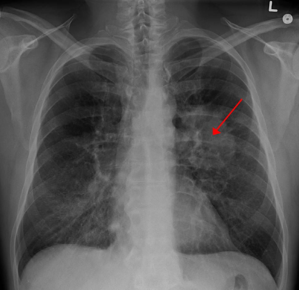
- Adenocarcinoma: peripheral; TTF-1 +, napsin A +; EGFR/ALK/ROS1/KRAS; associated with hypertrophic pulmonary osteoarthropathy (clubbing).
- Squamous cell: central, cavitary; keratin pearls + intercellular bridges; PTHrP → hypercalcemia.
- Small cell (neuroendocrine): central; Kulchitsky cells; chromogranin / synaptophysin / NSE +; paraneoplastic SIADH, ectopic ACTH, Lambert-Eaton; RB1/TP53 loss; almost always disseminated at diagnosis.
- Large cell: undifferentiated, highly anaplastic, poor prognosis; may secrete β-hCG → gynecomastia.
- Bronchial carcinoid: low-grade, well-differentiated neuroendocrine tumor; indolent with good prognosis. Carcinoid syndrome is uncommon and — unlike midgut/GI carcinoids — does not require liver metastases (pulmonary venous drainage bypasses hepatic first-pass metabolism).
Histologic Subtype Comparison
| Subtype | Location | Smoking link | Buzzwords / paraneoplastic |
|---|---|---|---|
| Adenocarcinoma | Peripheral | +/− (most common in never-smokers) | TTF-1+, napsin A+; EGFR/ALK; clubbing (HPOA) |
| Squamous cell | Central | Strong | Cavitation, keratin pearls; PTHrP → hyperCa |
| Small cell | Central | Strongest | Neuroendocrine; SIADH, ACTH, Lambert-Eaton |
| Large cell | Peripheral | + | Undifferentiated/anaplastic; β-hCG |
Vignette: A 63-year-old man with a 40-pack-year history has cough, weight loss, constipation, and confusion. Labs: Ca²⁺ 13.6, PTH low, PTHrP elevated, phosphate low. CXR shows a central cavitary mass.
- Diagnosis: Squamous cell carcinoma with PTHrP-mediated hypercalcemia (humoral hypercalcemia of malignancy). PTHrP mimics PTH → increased renal Ca²⁺ reabsorption and phosphate wasting, with suppressed native PTH.
- Next best step (dx): obtain tissue — a central lesion is best sampled by bronchoscopy/EBUS; a peripheral lesion by CT-guided biopsy.
- Next best step (tx): symptomatic hyperCa → aggressive IV isotonic saline first, then treat the tumor; add bisphosphonate/calcitonin for severe/persistent elevation.
- Squamous → PTHrP → hypercalcemia (low PTH, low phosphate).
- Small cell → SIADH → euvolemic hyponatremia.
- Small cell → ectopic ACTH → Cushing (HTN, hypokalemia, hyperglycemia; rapid onset, often without classic habitus).
- Small cell → Lambert-Eaton (anti–voltage-gated Ca²⁺ channel): proximal weakness that improves with repeated use ± autonomic sx — opposite of myasthenia gravis (worsens with use).
- Small cell → anti-Hu → paraneoplastic encephalomyelitis / subacute cerebellar degeneration.
- Large cell → β-hCG (gynecomastia).
- NSCLC/adeno → hypertrophic pulmonary osteoarthropathy (clubbing + periostitis).

Vignette: A 58-year-old smoker reports shoulder and medial-arm pain, hand-muscle wasting, and a drooping right eyelid. Exam: ptosis, miosis, and anhidrosis on the right.
- Diagnosis: Pancoast (superior sulcus) tumor invading the brachial plexus (C8–T1) and the cervical sympathetic chain → Horner syndrome; usually NSCLC.
- Also recognize: hoarseness = recurrent laryngeal nerve; facial/arm swelling + distended neck veins = SVC syndrome (classically small cell).
- Next best step: CT chest + MRI of the thoracic inlet/brachial plexus to define local invasion, then biopsy for tissue diagnosis and staging.
Screening, Nodules, Staging & PFTs
Screening (USPSTF 2021): annual low-dose CT for adults 50–80 yrs with a ≥20 pack-year history who currently smoke or quit within the past 15 years.
Solitary pulmonary nodule: always compare with old films. Benign clues: popcorn calcification (hamartoma), laminated/central calcification (granuloma), stability >2 yrs. Suspicious: spiculated, >2 cm, upper lobe, older heavy smoker → biopsy/PET.
Workup/staging: get tissue, then PET-CT + contrast CT chest; sample mediastinal nodes by EBUS; brain MRI (esp. SCLC & adeno). NSCLC → TNM (I–IV); SCLC → limited vs extensive.
Pre-resection PFTs: measure FEV₁ and DLCO and estimate predicted postoperative values — ppo-FEV₁ and ppo-DLCO >40% predicted generally signal acceptable operative risk (values <30–40% flag high risk → consider exercise testing).
SCLC vs NSCLC — Staging & Management
| Feature | SCLC | NSCLC |
|---|---|---|
| Frequency / behavior | ~15%; very aggressive, early mets | ~85%; comparatively slower |
| Staging | Limited vs extensive | TNM (I–IV) |
| Surgery? | Rarely (usually disseminated) | Yes for early stage (lobectomy) |
| Backbone therapy | Platinum + etoposide + RT; ± prophylactic cranial irradiation | Stage-based: surgery ± adjuvant chemo → chemoRT → systemic |
| Targeted / immuno | Add immunotherapy (extensive stage) | EGFR/ALK TKIs; PD-1/PD-L1 (pembrolizumab) |
SPHERE — regional complications of lung cancer:
- S — SVC syndrome (facial/arm swelling, distended neck veins)
- P — Pancoast tumor (superior sulcus)
- H — Horner syndrome (ptosis, miosis, anhidrosis)
- E — Endocrine paraneoplastic (PTHrP, SIADH, ACTH)
- R — Recurrent laryngeal nerve → hoarseness (also phrenic → diaphragm paralysis)
- E — Effusions (malignant pleural or pericardial)
Practice Pulmonary now
Board-style questions, spaced-repetition flashcards, and a Socratic AI tutor — free to start.