Lower Limb Nerve Injuries
A Step 1–focused walkthrough of the lumbosacral plexus that maps each lower-limb nerve to its roots, motor and sensory territory, classic injury mechanism, and signature deficit/gait, anchored by high-yield vignettes (foot drop, Trendelenburg, meralgia paresthetica) and a mechanism → nerve reverse-lookup. Two comparison tables and the genuine anatomy mnemonics (PED/TIP, \"sound side sags,\" NAVEL) tie it together.
Overview: The Lumbosacral Plexus Roadmap
Every lower-limb nerve arises from the lumbosacral plexus (L1–S4), and the boards test it as a set of predictable mechanism → deficit pairs. Split the plexus into two functional halves:
- Lumbar plexus (L1–L4) forms within the psoas major and supplies the anterior and medial thigh. Its two examinable motor nerves are the femoral (knee extension, hip flexion) and the obturator (thigh adduction).
- Sacral plexus (L4–S4) supplies the gluteal region, posterior thigh, and everything below the knee. Its key nerves are the superior gluteal, inferior gluteal, sciatic (which splits into the tibial and common fibular/peroneal), and the pudendal.
A useful organizing rule: nerves from the posterior divisions (femoral, superior gluteal, common fibular, deep fibular) supply knee extensors, dorsiflexors, and hip abductors, while anterior-division nerves (obturator, tibial) supply hip adductors, knee flexors, and plantarflexors. The one classic exception is hip flexion — the femoral nerve (a posterior-division nerve) still supplies iliacus. On exam day each nerve maps cleanly to a fracture site, body position, or injection error and yields a reproducible motor loss + sensory loss + gait.
- Femoral (L2–L4): knee extension (quadriceps) + hip flexion (iliopsoas); sensory to anterior thigh and medial leg/foot (saphenous branch). Lesion → weak knee extension + ↓ patellar reflex.
- Obturator (L2–L4): thigh adduction; sensory to medial thigh.
- Superior gluteal (L4–S1): hip abduction (gluteus medius/minimus, TFL). Lesion → Trendelenburg sign (pelvis drops on the side opposite the lesion when standing on the affected leg).
- Inferior gluteal (L5–S2): hip extension (gluteus maximus). Lesion → trouble climbing stairs / rising from a chair.
- Sciatic (L4–S3): hamstrings + all muscles below the knee (via tibial + common fibular).
- Common fibular / peroneal (L4–S2): dorsiflexion + eversion; sensory dorsum of foot + lateral leg. Lesion → FOOT DROP.
- Tibial (L4–S3): plantarflexion + inversion + toe flexion; sensory sole. Lesion → can't stand on tiptoe.
- Pudendal (S2–S4): perineum + external urethral/anal sphincters; blocked at the ischial spine.
- Lateral femoral cutaneous (L2–L3): purely sensory anterolateral thigh → meralgia paresthetica.
- Femoral triangle contents lateral→medial = NAVEL; the femoral nerve lies OUTSIDE the femoral sheath.
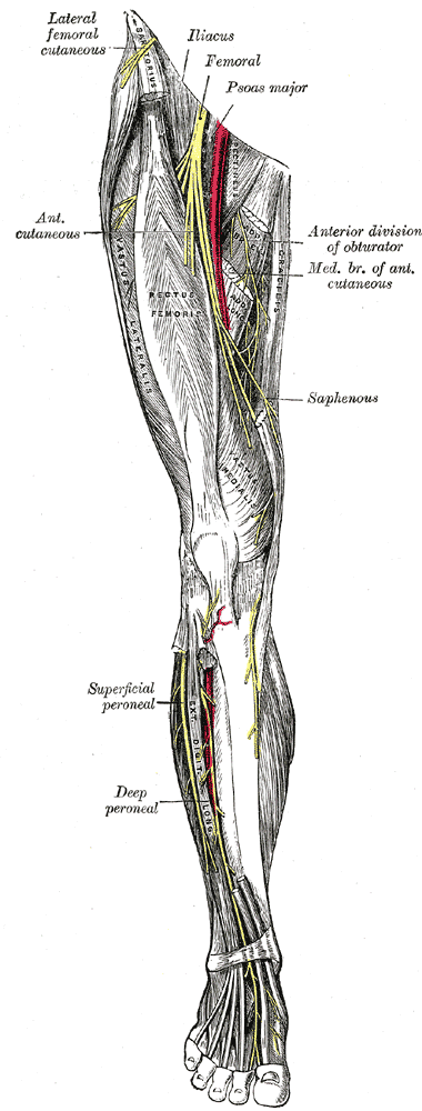
Master Comparison: Nerve → Roots → Motor → Sensory → Injury
| Nerve | Roots | Motor lost | Sensory lost | Classic cause | Signature deficit / gait |
|---|---|---|---|---|---|
| Femoral | L2–L4 | Knee extension (quadriceps), hip flexion (iliopsoas) | Anterior thigh, medial leg (saphenous) | Pelvic fracture, retroperitoneal/iliacus hematoma, lithotomy position, hip surgery | Buckling knee, ↓ patellar reflex |
| Obturator | L2–L4 | Thigh adduction | Medial thigh | Anterior hip dislocation, pelvic surgery (e.g., node dissection), obturator hernia | Weak adduction, leg swings laterally |
| Superior gluteal | L4–S1 | Hip abduction (glut. medius/minimus, TFL) | — | Misplaced IM injection (superomedial quadrant), hip surgery, posterior hip dislocation | Trendelenburg gait (contralateral pelvic drop) |
| Inferior gluteal | L5–S2 | Hip extension (glut. maximus) | — | Posterior hip dislocation, pelvic surgery | Can't climb stairs / rise from seat; gluteal lurch |
| Sciatic | L4–S3 | Hamstrings + all below knee | Posterolateral leg + foot (spares saphenous) | Posterior hip dislocation, deep IM injection, disc herniation | Weak knee flexion, flail foot |
| Common fibular (peroneal) | L4–S2 | Dorsiflexion + eversion (+ toe extension) | Dorsum of foot, lateral leg | Fibular neck fracture, tight cast, leg crossing | Foot drop, steppage gait; foot inverted + plantarflexed |
| Tibial | L4–S3 | Plantarflexion, inversion, toe flexion | Sole of foot | Popliteal injury (knee dislocation, Baker cyst), tarsal tunnel | Can't stand on tiptoe; loss of inversion |
| Pudendal | S2–S4 | External urethral/anal sphincters | Perineum, genitalia | Childbirth stretch, prolonged cycling | Incontinence, perineal numbness |

Femoral nerve — An anticoagulated patient (or hemophiliac) develops groin pain, then cannot extend the knee, has a diminished patellar reflex, and reports numbness over the anterior thigh and medial leg. → Femoral nerve compressed by a retroperitoneal / iliacus hematoma. Also classic after lithotomy positioning (childbirth, pelvic surgery) or pelvic fracture.
Obturator nerve — After a difficult forceps delivery or pelvic mass resection, a patient has weak thigh adduction and numbness over the medial thigh. → Obturator nerve (also anterior hip dislocation, obturator hernia).
Superior gluteal nerve — A patient receives an intramuscular injection in the superomedial gluteal quadrant. When they stand on that leg, the opposite side of the pelvis drops — a positive Trendelenburg sign. → Superior gluteal nerve. Prevention: inject the superolateral (upper-outer) quadrant.
Inferior gluteal nerve — After a posterior hip dislocation, a patient has difficulty standing up from a chair and climbing stairs but a near-normal gait on level ground. → Inferior gluteal nerve (gluteus maximus).
Common fibular (peroneal) nerve — A patient falls asleep with legs crossed, or has a lower-leg cast, or is struck on the lateral knee, and develops foot drop: cannot dorsiflex or evert, the foot hangs inverted and plantarflexed, and there is numbness over the dorsum of the foot. Walking shows a high-stepping (steppage) gait. The nerve wraps the fibular neck superficially — the most commonly injured nerve of the lower limb. Remember: P-E-D (Peroneal Everts, Dorsiflexes) → foot droP-P-E-D.
Sciatic nerve — Dashboard trauma drives the flexed, adducted hip posteriorly (posterior hip dislocation), stretching the sciatic nerve — its common fibular division is preferentially injured, so foot drop dominates. Also from deep IM injection in the wrong gluteal quadrant or disc herniation.
Tibial nerve — A patient with a posterior knee dislocation, a Baker cyst, or tarsal tunnel syndrome (compression behind/below the medial malleolus) cannot stand on tiptoe, has weak inversion and toe flexion, and reports tingling/numbness over the sole. → Tibial nerve.
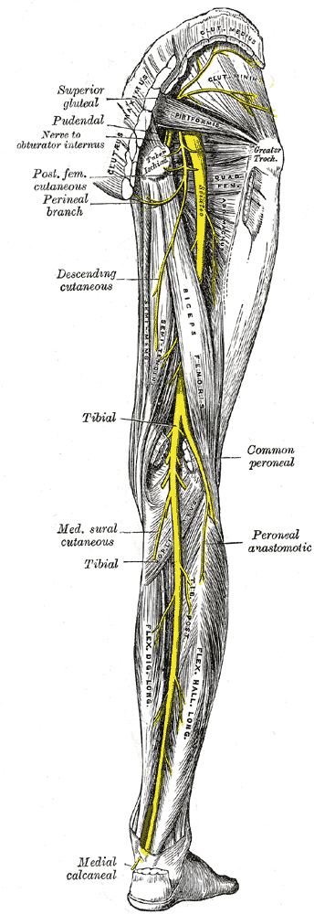
- PED = common Peroneal (fibular) Everts and Dorsiflexes. Lose it and the foot is droPPED (foot drop, sensory loss over the dorsum).
- TIP = Tibial Inverts and Plantarflexes (sensory loss over the sole).
- Trendelenburg: "the sound side sags." With a superior gluteal lesion, the healthy (contralateral) side of the pelvis drops when weight is on the affected leg.
- IM injections → superolateral quadrant of the buttock, sparing the sciatic (lower quadrants) and the superior gluteal (superomedial).
- Femoral triangle = NAVEL (lateral → medial): Nerve, Artery, Vein, Empty space (femoral canal), Lymphatics. Only the Nerve is outside the femoral sheath.
- Obturator and Femoral are both "2, 3, 4" (L2–L4).
Gait Patterns Decoded
| Gait | Nerve / muscle | Mechanism | Key exam finding |
|---|---|---|---|
| Trendelenburg | Superior gluteal (glut. medius/minimus) | Loss of hip-abductor pelvic stabilization | Pelvis drops on the contralateral (swing) side; trunk lists toward the affected side to compensate |
| Gluteus maximus lurch | Inferior gluteal (glut. maximus) | Loss of hip extension | Posterior trunk thrust at heel strike; trouble on stairs |
| Steppage (high-stepping) | Common fibular (foot drop) | Loss of dorsiflexion | Exaggerated hip + knee flexion to clear the toes; audible foot slap |
| Waddling (bilateral) | Bilateral hip-abductor weakness | Often proximal myopathy (e.g., muscular dystrophy) — contrast with a unilateral nerve lesion | Side-to-side sway; a bilateral Trendelenburg |
Meralgia paresthetica — An obese patient (or one who is pregnant, wears a tight belt, or carries a heavy tool belt) reports burning, tingling, and numbness over the anterolateral thigh with no motor weakness and normal reflexes. → Entrapment of the lateral femoral cutaneous nerve (L2–L3) under the inguinal ligament near the ASIS. Purely sensory = no weakness is the giveaway.
Pudendal nerve — For anesthesia during the second stage of labor, a pudendal nerve block is placed at the ischial spine (palpated via the sacrospinous ligament). Chronic perineal numbness in a competitive cyclist ('cyclist's syndrome') reflects pudendal neuralgia.
- Foot drop / fibular neck fracture / crossed legs / tight leg cast → common fibular (peroneal)
- Trendelenburg / superomedial IM injection / hip surgery → superior gluteal
- Can't climb stairs or rise from a chair → inferior gluteal
- Retroperitoneal (iliacus) hematoma / lithotomy / ↓ knee jerk → femoral
- Anterior hip dislocation / obturator hernia / weak adduction → obturator
- Dashboard posterior hip dislocation → sciatic (± its common fibular part)
- Knee dislocation / tarsal tunnel / can't tiptoe → tibial
- Anterolateral thigh burning, no weakness → lateral femoral cutaneous (meralgia paresthetica)
- Ischial spine block during delivery → pudendal
Practice Anatomy now
Board-style questions, spaced-repetition flashcards, and a Socratic AI tutor — free to start.