Laryngeal Disorders & Hoarseness
A high-yield Step 2 CK walkthrough of hoarseness: when to scope (≥2–3 weeks or red flags), the benign vocal-fold lesions, vocal-cord paralysis with RLN anatomy, and laryngeal SCC — built around next-best-step decisions and classic vignette buzzwords.
Approach to Hoarseness
Hoarseness (dysphonia) is an altered voice quality from anything that disrupts smooth vocal-fold vibration. Most acute cases are self-limited viral laryngitis. The board-defining rule: hoarseness lasting ≥2–3 weeks, or any red-flag feature, requires direct visualization by flexible laryngoscopy — chiefly to exclude squamous cell carcinoma (SCC) in the older smoker/drinker.
Voice is generated as the vocal folds adduct and vibrate. Nearly all intrinsic laryngeal muscles are supplied by the recurrent laryngeal nerve (RLN), a branch of the vagus. The left RLN loops under the aortic arch, giving it a long intrathoracic course vulnerable to compression by lung/mediastinal tumor or aneurysm. Organize causes into four buckets: benign mucosal lesions (nodules, polyps, Reinke edema, papillomas), neuromuscular (cord paralysis), inflammatory (laryngitis, reflux), and malignant (SCC).

- Acute hoarseness (<2 wk) with URI symptoms → viral laryngitis; voice rest and hydration; antibiotics not indicated
- Hoarseness ≥2–3 weeks → flexible laryngoscopy to inspect the folds (next best step)
- Red flags = tobacco/heavy alcohol, neck mass, dysphagia/odynophagia, hemoptysis, referred otalgia, stridor, weight loss → urgent scope ± imaging
- Vocal cord nodules (bilateral, voice overuse — teachers, singers, children) → voice/speech therapy first-line, not surgery
- Vocal polyp / Reinke edema (smoking) → smoking cessation + microlaryngeal excision
- Laryngopharyngeal reflux (LPR): morning hoarseness, throat clearing, globus, posterior laryngeal edema → PPI + lifestyle
- Isolated left vocal-cord paralysis with no laryngeal cause → CT neck/chest to hunt tumor along the RLN
- Bilateral cord paralysis (classically post-thyroidectomy) → inspiratory stridor and airway emergency
Benign Vocal-Fold Lesions
| Lesion | Key features (buzzword) | Cause | First-line management |
|---|---|---|---|
| Vocal nodules | Bilateral, symmetric; anterior–middle third; teacher/singer/child | Chronic voice overuse | Voice therapy |
| Vocal polyp | Usually unilateral, pedunculated | Smoking; single phonotrauma | Stop smoking; excision |
| Reinke edema | Bilateral floppy "polypoid" folds; low, gruff voice | Smoking (± LPR, hypothyroid) | Stop smoking; surgery |
| Contact granuloma/ulcer | Posterior larynx (arytenoid) | GERD/LPR, intubation, throat clearing | Treat reflux; voice rest |
| Papilloma (RRP) | Wart-like; child or young adult | HPV 6 & 11 | Surgical debulking (recurs) |
Vignette: A 34-year-old elementary-school teacher who also sings in a choir reports 4 months of a raspy voice and vocal fatigue, worse by day's end and after loud events, better with rest. She does not smoke; no dysphagia or weight loss. Laryngoscopy shows symmetric whitish swellings at the junction of the anterior and middle thirds of both vocal folds.
Diagnosis: Vocal cord nodules ("singer's/screamer's nodules") — bilateral, from chronic voice overuse.
Next best step / management: Voice (speech) therapy with behavioral modification — first-line and frequently curative; surgery is reserved for refractory lesions. The bilateral, symmetric location and overuse history separate nodules from a (typically unilateral) polyp.
Vignette: A 63-year-old man with a 40-pack-year smoking history and daily alcohol use has 7 weeks of progressive hoarseness, now with right otalgia and a firm neck node. Flexible laryngoscopy reveals an irregular exophytic mass on the true vocal cord.
Diagnosis: Glottic squamous cell carcinoma. Glottic tumors cause hoarseness early (often caught at an earlier stage); referred ear pain travels via the vagus (CN X, auricular/Arnold branch).
Next best step: Direct laryngoscopy with biopsy for tissue diagnosis, plus CT neck/chest for staging. Risk factors for laryngeal SCC: tobacco + alcohol (synergistic) — HPV is the classic driver of oropharyngeal (tonsil/base-of-tongue) SCC, not glottic cancer. Contrast: supraglottic cancers have rich lymphatics and present later with dysphagia/neck mass.
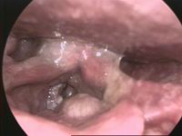
Vignette: A 58-year-old smoker has a breathy, weak voice and a weak, "bovine" cough (poor glottic closure). Laryngoscopy shows the left vocal cord fixed in a paramedian position; chest imaging shows a left hilar mass.
Diagnosis: Left vocal-cord paralysis from recurrent laryngeal nerve invasion by lung cancer (the left RLN loops under the aortic arch through the aortopulmonary window).
Next best step: biopsy the mass; image the entire RLN course (skull base → aortopulmonary window) when no laryngeal cause is seen. Teaching points: unilateral RLN palsy → breathy hoarseness (opposite cord compensates over time); bilateral (post-thyroidectomy) → cords near midline → stridor/airway compromise. Ortner (cardiovocal) syndrome = left RLN compressed by cardiovascular enlargement (classically a dilated left atrium from mitral stenosis; also aortic/pulmonary artery dilation).
PCA — "Please Come Apart"
- The Posterior CricoArytenoid is the only abductor — it opens the glottis for breathing (protects the airway). Every other intrinsic muscle either adducts (closes) the folds (lateral cricoarytenoid, interarytenoids) or tenses them (cricothyroid, thyroarytenoid) — the PCA alone abducts.
"All RLN except the cricothyroid"
- All intrinsic laryngeal muscles are innervated by the recurrent laryngeal nerve, EXCEPT the cricothyroid, supplied by the external branch of the superior laryngeal nerve (SLN); it tenses/elongates the cord to raise pitch.
- SLN injury (e.g., thyroidectomy) → subtle voice fatigue, loss of high notes; RLN injury → frank hoarseness or, if bilateral, airway compromise.
Practice ENT (Otolaryngology) now
Board-style questions, spaced-repetition flashcards, and a Socratic AI tutor — free to start.