Interstitial Lung Disease & Pneumoconioses
A Step 1 high-yield lesson on interstitial lung disease and the pneumoconioses, moving from restrictive pathophysiology through PFT/imaging/lab diagnosis to management, anchored by classic vignette buzzwords, a UIP/IPF focus, and next-best-step decisions. Includes comparison tables (pneumoconioses; obstructive vs restrictive PFTs) and two verified Wikimedia figures.
What ILD Is and Why It Restricts
Interstitial lung diseases (ILDs) are a heterogeneous group of >200 disorders that scar and thicken the alveolar–capillary interstitium, producing a restrictive ventilatory defect. Chronic injury to type I/II pneumocytes and endothelium triggers aberrant fibroblast activation and collagen deposition, stiffening the lung. The result is low compliance, reduced lung volumes, and impaired gas diffusion.
Patients present with insidious exertional dyspnea and a dry cough, fine end-inspiratory "Velcro" crackles at the bases, and digital clubbing (especially IPF and asbestosis); late disease brings hypoxemia and cor pulmonale.
Boards group ILD by cause: idiopathic (IPF, NSIP), occupational pneumoconioses (asbestos, silica, coal, beryllium), granulomatous (sarcoidosis, hypersensitivity pneumonitis), connective-tissue-disease-associated, and drug/radiation-induced. The unifying testable signature is a restrictive pattern with a preserved or elevated FEV1/FVC ratio plus a reduced DLCO.
- PFTs (restrictive): ↓FVC, ↓TLC, ↓RV; FEV1/FVC normal or ↑ (≥0.70); ↓DLCO from loss of gas-exchange surface.
- Flow-volume loop: small and narrow, shifted rightward toward low lung volumes; the scooped/coved expiratory limb of obstruction is absent.
- Exam: bibasilar dry "Velcro" crackles, clubbing; later loud P2, JVD, edema (cor pulmonale).
- HRCT is the key noninvasive test. Honeycombing + traction bronchiectasis + subpleural, basal reticulation = UIP pattern (classic for IPF).
- Upper-lobe predominance: silicosis, coal, berylliosis, sarcoidosis, hypersensitivity pneumonitis, TB.
- Lower-lobe predominance: IPF, asbestosis, connective-tissue disease, most drug-induced.
- Drug causes to memorize: bleomycin, busulfan, amiodarone, methotrexate, nitrofurantoin; plus radiation.
A 62-year-old man reports 2 years of progressive exertional dyspnea and dry cough. In his 30s he installed pipe insulation in a naval shipyard. Exam: fine bibasilar Velcro crackles and clubbing. CXR shows lower-lobe reticular opacities and calcified pleural plaques along the diaphragm.
- Diagnosis: Asbestosis (latency 20–40 yrs). Biopsy shows golden-brown, dumbbell-shaped ferruginous (asbestos) bodies.
- Pleural plaques mark exposure but are benign and do NOT transform into mesothelioma.
- Highest-yield association: the greatest absolute cancer risk is bronchogenic carcinoma (synergistic with smoking); mesothelioma is the most specific, smoking-independent tumor.
- Next best step if a new bloody, exudative pleural effusion appears → suspect mesothelioma and obtain pleural biopsy (fluid cytology alone is often insufficient).
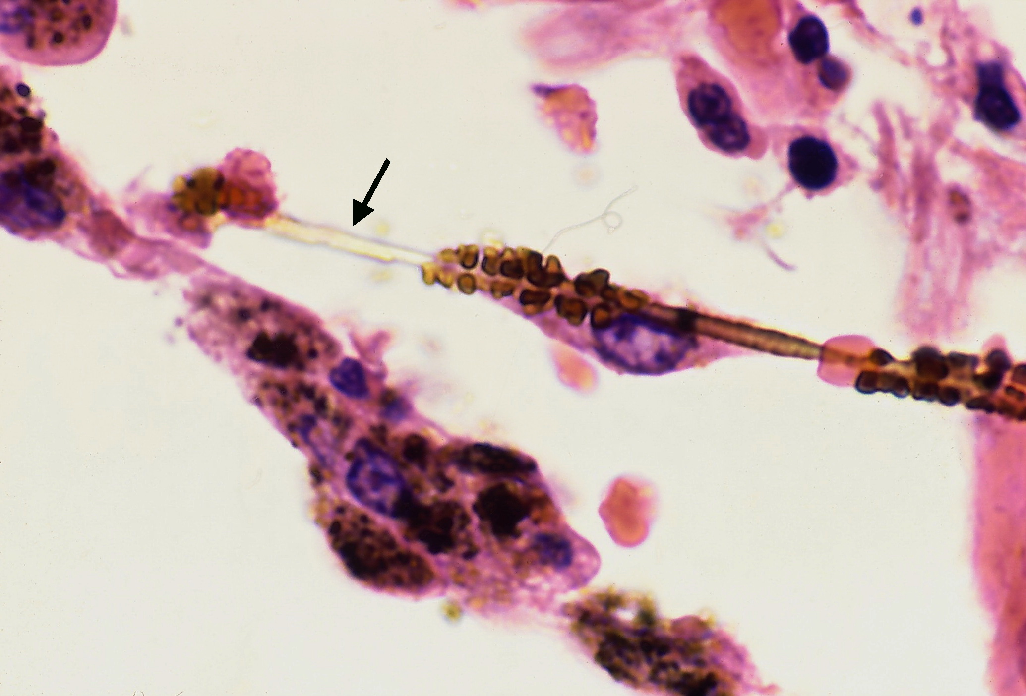
Pneumoconioses at a Glance
| Disease | Exposure | Lobe | Classic clues | Complication |
|---|---|---|---|---|
| Asbestosis | Shipyards, insulation, roofing, plumbing | Lower | Calcified pleural plaques; ferruginous bodies | Bronchogenic CA (most common) > mesothelioma |
| Silicosis | Sandblasting, mining, quarrying, foundry | Upper | "Eggshell" hilar node calcification; silicotic nodules | ↑TB (impaired macrophage killing); lung CA; PMF |
| Coal worker's | Coal mining | Upper | Coal macules, anthracosis; Caplan (+RA) | Progressive massive fibrosis; no independent ↑ lung CA |
| Berylliosis | Aerospace, electronics, ceramics, nuclear | Upper | Non-caseating granulomas (mimics sarcoid); hilar adenopathy | Steroid-responsive; ↑ lung CA |
Idiopathic Pulmonary Fibrosis (IPF)
IPF is the most common idiopathic interstitial pneumonia—typically a man >60, often a former smoker, with progressive dyspnea and a UIP pattern. HRCT shows subpleural, basal-predominant reticulation with honeycombing and traction bronchiectasis; histology shows spatial and temporal heterogeneity with fibroblastic foci (patchy dense fibrosis beside normal lung).
When the HRCT UIP pattern is definite, diagnosis is made without biopsy—but first exclude mimics (asbestos exposure, connective-tissue disease, drugs, hypersensitivity pneumonitis).
Management:
- Corticosteroids do NOT help and may harm (unlike sarcoid/HP/NSIP)—the triple therapy of prednisone/azathioprine/N-acetylcysteine increased mortality in trials.
- Antifibrotics—pirfenidone or nintedanib—slow FVC decline but do not reverse fibrosis.
- Lung transplantation is the only definitive therapy.
- Supportive: O2, pulmonary rehab, vaccination.
Median survival ~3–5 years; acute exacerbations and cor pulmonale drive mortality.

- Sarcoidosis: non-caseating granulomas; bilateral hilar lymphadenopathy; classically young African-American women; ↑ACE; hypercalcemia (macrophage 1α-hydroxylase → ↑1,25-vitamin D); erythema nodosum, uveitis; asteroid & Schaumann bodies. Many resolve spontaneously; symptomatic disease → steroids.
- Hypersensitivity pneumonitis: mixed type III/IV reaction to an inhaled antigen—Farmer's lung (thermophilic actinomycetes), bird-fancier's lung; upper-lobe; improves with antigen avoidance ± steroids.
- Berylliosis is histologically indistinguishable from sarcoid—ask about occupation (aerospace/electronics) and confirm with the beryllium lymphocyte proliferation test (BeLPT).
- Key contrast: steroids help sarcoid/HP/berylliosis but not IPF.
Where fibrosis sits narrows the differential. Upper-lobe predominance = CHARTS:
- C — Coal worker's pneumoconiosis
- H — Hypersensitivity pneumonitis / Histiocytosis (Langerhans)
- A — Ankylosing spondylitis
- R — Radiation
- T — Tuberculosis
- S — Silicosis / Sarcoidosis
Everything else favors the lower lobes—IPF, asbestosis, connective-tissue disease, and most drug-induced fibrosis.
Quick hook: "Asbestos drags you down" (lower zones). The mineral-dust pneumoconioses (silica, coal, beryllium) and granulomatous ILDs (sarcoid, HP) cluster in the upper zones, attributed to relatively poorer lymphatic clearance there.
A 48-year-old sandblaster has 3 years of dyspnea. CXR shows small upper-lobe nodules and "eggshell" calcification of hilar lymph nodes. He now reports weight loss, night sweats, and hemoptysis.
- Diagnosis: Silicosis, now complicated by reactivation tuberculosis—silica is toxic to alveolar macrophages and impairs intracellular killing of M. tuberculosis ("silicotuberculosis").
- Next best step: because he has symptoms of active disease, obtain sputum AFB smear, culture, and NAAT (± chest imaging) and treat active TB once confirmed. IGRA/PPD detect infection and can be falsely negative in active TB, so they cannot exclude it.
- Boards nuance: a silicosis patient with a positive IGRA/PPD but no active disease still warrants latent-TB therapy because reactivation risk is high.
- Silica independently raises lung cancer risk (IARC group 1 carcinogen); large coalescing upper-lobe masses = progressive massive fibrosis.
Contrast trap: upper-lobe nodules + hilar adenopathy can mimic sarcoid—but the occupational history, eggshell nodes, and TB symptoms point to silicosis.
Obstructive vs Restrictive PFTs
| Parameter | Obstructive (COPD/asthma) | Restrictive (ILD) |
|---|---|---|
| FEV1 | ↓↓ | ↓ |
| FVC | ↓ or normal | ↓↓ |
| FEV1/FVC | ↓ (<0.70) | normal or ↑ (≥0.70) |
| TLC | ↑ (air trapping) | ↓ |
| DLCO | ↓ emphysema; normal asthma | ↓ in ILD; normal/↑ in chest-wall or neuromuscular |
| Flow-volume loop | scooped/coved expiratory limb | small, narrow, right-shifted |
Practice Pulmonary now
Board-style questions, spaced-repetition flashcards, and a Socratic AI tutor — free to start.