Skin & Integument Histology
A board-focused tour of skin histology: the five epidermal layers and their resident cells, dermal receptors and glands, and the two high-yield blistering diseases (pemphigus vulgaris vs bullous pemphigoid) defined by the junction protein targeted and the level of the split.
Skin: Architecture & Why Boards Test It
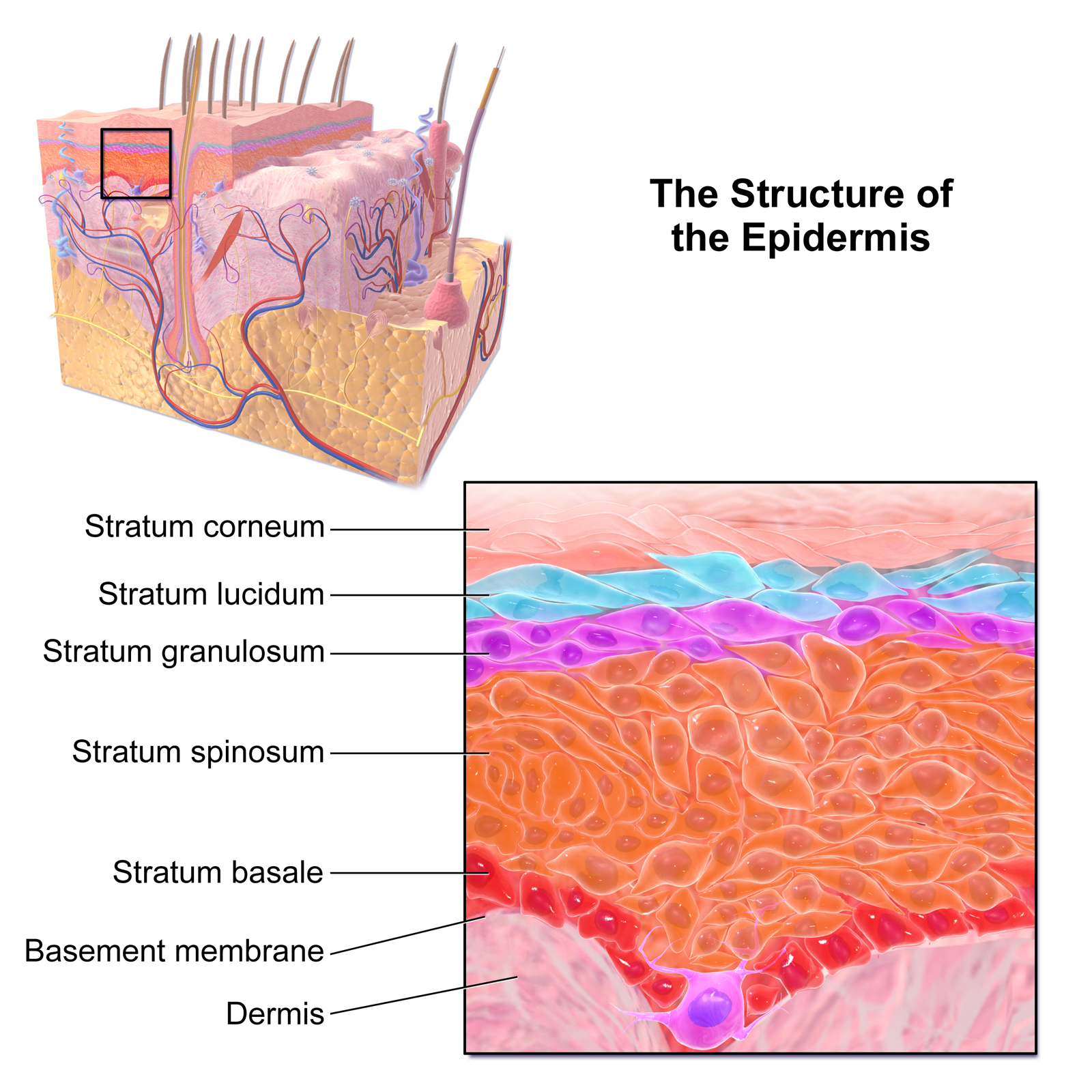
Skin is the body's largest organ, built in three layers: the epidermis (keratinized stratified squamous epithelium, ectoderm-derived), the dermis (connective tissue), and the hypodermis (subcutaneous fat). The epidermis is avascular and renews from the stratum basale, whose keratinocytes migrate up, differentiate, and die to form the protective stratum corneum — roughly a 4-week journey. Besides keratinocytes, the epidermis holds melanocytes (pigment), Langerhans cells (immune surveillance), and Merkel cells (touch).
Step 1 tests skin histology mostly through blistering diseases — where the level of the split and the junction protein targeted set the diagnosis — and through the origins and immunostains of the resident non-keratinocyte cells. Know the layers cold, then map each disease onto them.
- Layers (deep → superficial): basale → spinosum → granulosum → lucidum → corneum
- Stratum basale: single row of mitotic stem cells; melanocytes and Merkel cells live here
- Stratum spinosum: "spiny" desmosome-linked keratinocytes; Langerhans cells reside here
- Stratum granulosum: keratohyalin granules; keratinocytes lose nuclei
- Stratum lucidum: present only in thick skin — palms and soles
- Stratum corneum: anucleate keratin flakes; thickened in psoriasis and calluses
- Keratinocytes = the majority cell; joined to each other by desmosomes and to the basement membrane by hemidesmosomes
- Renewal is bottom-up: mitosis in basale, terminal differentiation in corneum
"Come, Let's Get Sun Burned" — epidermal layers, surface → deep:
- Corneum, Lucidum, Granulosum, Spinosum, Basale
Blister level:
- Bullous pemphigoid = Below the epidermis (subepidermal); targets the hemidesmosome (BP180/BP230); Nikolsky negative; more benign
- Pemphigus vulgaris = higher, intraepidermal (suprabasal); targets desmoglein 1/3; Nikolsky positive; potentially fatal
desmoGlein → pemphiGus — both carry a G, and pemphigus is the one that targets desmoglein.
Pemphigus Vulgaris vs Bullous Pemphigoid
| Feature | Pemphigus vulgaris | Bullous pemphigoid |
|---|---|---|
| Target antigen | Desmoglein 1 & 3 (desmosome) | BP180 / BP230 (hemidesmosome) |
| Split level | Intraepidermal (suprabasal) | Subepidermal |
| Bullae | Flaccid, rupture easily | Tense, stay intact |
| Nikolsky sign | Positive | Negative |
| Oral mucosa | Commonly involved (often first) | Usually spared |
| DIF pattern | Intercellular IgG, "net/fishnet" | Linear IgG + C3 at BMZ |
| Histology clue | "Row of tombstones", acantholysis | Eosinophil-rich subepidermal blister |
| Typical patient | Middle-aged; more severe | Elderly; milder course |
Vignette: A 50-year-old has painful flaccid bullae and erosions on the trunk plus oral ulcers that appeared before the skin lesions. Lateral pressure on normal-looking skin extends the blister (Nikolsky positive). Biopsy shows suprabasal acantholysis with basal cells still anchored to the basement membrane ("row of tombstones"); direct immunofluorescence shows intercellular IgG in a net/fishnet pattern.
Diagnosis: Pemphigus vulgaris — autoantibodies vs desmoglein 1/3.
Next best step: Confirm with skin biopsy + direct immunofluorescence (plus serum anti-desmoglein ELISA), then start systemic corticosteroids, adding rituximab as first-line steroid-sparing therapy for moderate-to-severe disease. Untreated PV can be fatal from fluid loss and secondary infection.
- Papillary dermis: loose (areolar) CT; houses Meissner corpuscles — light/discriminative touch, in dermal papillae of glabrous skin
- Reticular dermis: dense irregular CT; deeper dermis/subcutis houses Pacinian corpuscles — deep pressure/vibration
- Merkel discs: static/light touch, slowly adapting (in stratum basale)
- Desmosome (desmoglein): keratinocyte–keratinocyte → target in pemphigus vulgaris
- Hemidesmosome (integrins / BP antigens): keratinocyte–basement membrane → target in bullous pemphigoid
- Glands: sebaceous = holocrine (whole cell disintegrates); eccrine sweat = merocrine, thermoregulation; apocrine = axilla/groin, body odor
- Epidermis is ectoderm-derived; melanocytes are neural crest-derived

Vignette: An 80-year-old develops large tense bullae on flexural surfaces and trunk; blisters do not rupture with lateral pressure (Nikolsky negative), and the oral mucosa is spared. Biopsy shows a subepidermal blister rich in eosinophils; direct immunofluorescence reveals a linear band of IgG and C3 along the basement membrane.
Diagnosis: Bullous pemphigoid — autoantibodies vs hemidesmosome proteins BP180 / BP230.
Next best step: Skin biopsy + direct immunofluorescence to separate it from pemphigus; treat with high-potency topical corticosteroids (e.g., clobetasol) for limited disease or systemic corticosteroids for extensive disease. Chronic but generally less life-threatening than pemphigus vulgaris.
- Melanocytes: neural crest origin; sit in stratum basale; make melanin via tyrosinase in melanosomes, then transfer it to keratinocytes
- Albinism: normal melanocyte number but defective tyrosinase / tyrosine transport → no pigment; ↑ skin cancer and ocular defects
- Vitiligo: autoimmune destruction of melanocytes → sharply demarcated depigmented patches (fewer melanocytes)
- Langerhans cells: dendritic antigen-presenting cells in stratum spinosum; bone-marrow/monocyte origin; CD1a+, S100+, langerin (CD207)+; contain Birbeck granules ("tennis-racket" on EM)
- Langerhans cell histiocytosis: clonal proliferation of these cells → lytic bone lesions and skin rash; Birbeck granules are diagnostic
- Merkel cells: basale mechanoreceptors; Merkel cell carcinoma is linked to Merkel cell polyomavirus and is aggressive
Practice Histology now
Board-style questions, spaced-repetition flashcards, and a Socratic AI tutor — free to start.