Innate vs Adaptive Immunity
A Step 1 high-yield lesson contrasting innate immunity (fast, non-specific, no memory: phagocytes, NK cells, complement, TLRs) with adaptive immunity (slower, antigen-specific, memory: T and B cells), then linking each key cell/molecule to its classic immunodeficiency and infection pattern. Includes comparison and defect-to-disease tables, innate and adaptive clinical vignettes, and standard board mnemonics. Reviewed and signed off as accurate.
Two arms, one system
The immune system defends through two integrated arms. Innate immunity is the fast (minutes-to-hours), germline-encoded, non-specific first line: physical barriers, phagocytes (neutrophils, macrophages), NK cells, complement, and pattern-recognition receptors (PRRs) such as Toll-like receptors that sense conserved microbial motifs (PAMPs). It has no immunologic memory — the response is essentially identical on every exposure.
Adaptive immunity is slower on first contact (days), exquisitely antigen-specific, and uniquely generates memory, so the second exposure is faster and stronger. It comprises T lymphocytes (cell-mediated) and B lymphocytes/plasma cells (humoral antibody). Receptor diversity is created somatically by VDJ recombination, not encoded in the germline.
The two arms are physically linked by the dendritic cell — the professional antigen-presenting cell that ingests microbes via innate PRRs, then migrates to lymph nodes and presents peptide on MHC to prime naive T cells. Innate signals (which TLRs fire, which cytokines are released) instruct the type of adaptive response that follows.

- Innate = fast, fixed, no memory. Same magnitude on repeat exposure; receptors are germline-encoded PRRs (e.g., TLRs).
- Adaptive = slow first time, memory-driven, antigen-specific. Receptor diversity via VDJ recombination; anamnestic (faster/stronger) secondary response.
- Innate cells: neutrophils, macrophages, dendritic cells, NK cells, mast cells, basophils, eosinophils — plus complement and PRRs.
- Adaptive cells: T cells (cell-mediated) and B cells → plasma cells (humoral antibody).
- Dendritic cell = the bridge: professional APC linking innate sensing to adaptive priming.
- MHC I (all nucleated cells + platelets; not RBCs) → presents endogenous peptides to CD8⁺ T cells. MHC II (professional APCs only) → presents exogenous peptides to CD4⁺ T cells.
- NK cells are innate lymphocytes: they kill "missing self" — cells that down-regulate MHC I (viruses, tumors) — and are not MHC-restricted.
- Complement, phagocytes, and NK cells act within hours; specific antibody and effector T cells take ~5–7 days on a primary response.
Innate vs adaptive at a glance
| Feature | Innate immunity | Adaptive immunity |
|---|---|---|
| Speed | Immediate (minutes–hours) | Delayed 1st exposure (~5–7 days) |
| Specificity | Broad, for conserved PAMPs | Highly specific for one epitope |
| Memory | None | Yes (faster, stronger 2nd time) |
| Receptors | Germline-encoded PRRs (TLRs) | Somatically rearranged (VDJ) BCR/TCR |
| Diversity | Limited, fixed | Enormous |
| Key cells | Neutrophils, macrophages, DCs, NK | T cells, B cells/plasma cells |
| Soluble effectors | Complement, lysozyme, cytokines | Antibodies |
| MHC restriction | No (NK sense "missing self") | Yes (CD8→MHC I, CD4→MHC II) |
Toll-like receptors (PRRs) → NF-κB → inflammatory cytokines:
- TLR4 → LPS (gram-negative endotoxin; uses CD14 co-receptor)
- TLR2 → peptidoglycan / lipoteichoic acid (gram-positive)
- TLR5 → flagellin
- TLR3 → dsRNA · TLR7/8 → ssRNA · TLR9 → unmethylated CpG DNA
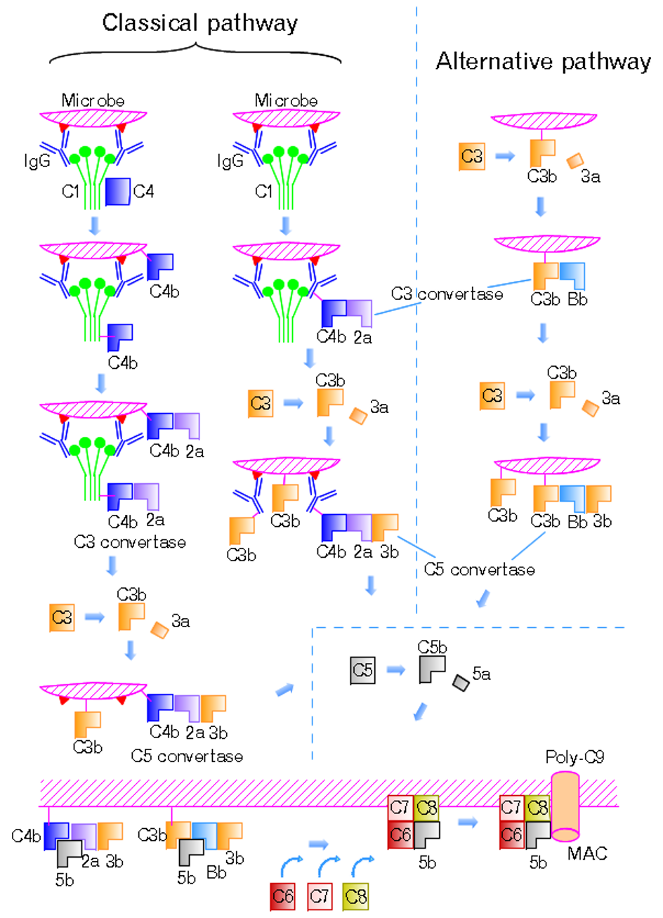
Complement (innate humoral):
- C3b = opsonin (with IgG). C3a, C4a, C5a = anaphylatoxins; C5a also = neutrophil chemotaxis.
- C5b–C9 = membrane attack complex (MAC) → lyses gram-negatives, especially Neisseria.
- Activation: classical (IgG/IgM–antigen, C1), alternative (microbial surfaces), lectin (MBL binds mannose).
NK cells: kill via perforin/granzyme; activated by IL-12, IL-15, IFN-α/β; mediate ADCC through CD16 (binds IgG Fc); markers CD16, CD56.
Key innate cytokines: IL-1, IL-6, TNF-α (fever/acute inflammation); IL-8 (neutrophil chemotaxis); IL-12 (drives Th1 + NK); IFN-γ (activates macrophages); IFN-α/β (type I interferons = antiviral).
Cell / molecule → function → defect
| Cell / molecule | Function | Defect → disease |
|---|---|---|
| NADPH oxidase (neutrophil) | Respiratory burst (O₂⁻ → H₂O₂) for killing | Chronic granulomatous disease — catalase⁺ infections; abnormal DHR/NBT |
| β2-integrin (CD18/LFA-1) | Leukocyte adhesion & extravasation | LAD type 1 — delayed cord separation, no pus |
| LYST | Phagolysosome fusion, vesicle trafficking | Chédiak-Higashi — giant granules, albinism |
| C5–C9 (MAC) | Lyse gram-negatives | Terminal complement deficiency — recurrent Neisseria |
| C1 esterase inhibitor | Regulates complement & bradykinin | Hereditary angioedema (↑ bradykinin) |
| CD55/CD59 (GPI-anchored) | Block complement on host cells | PNH — complement-mediated hemolysis |
| BTK | B-cell maturation | X-linked agammaglobulinemia |
| CD40L (on Th cells) | Ig class-switching | Hyper-IgM syndrome |
| Thymus (3rd/4th pouches) | T-cell maturation | DiGeorge (22q11.2) |
| IL-12 / IFN-γ axis | Th1 + macrophage activation | Disseminated mycobacterial disease |
| STAT3 | Th17 differentiation | Hyper-IgE (Job) syndrome |
1. Chronic granulomatous disease (NADPH oxidase). A 4-year-old boy has recurrent S. aureus skin abscesses, cervical lymphadenitis, and Aspergillus pneumonia. Flow cytometry dihydrorhodamine (DHR) test is abnormal (impaired oxidative burst). → Susceptibility to catalase-positive organisms (they degrade their own H₂O₂, and the neutrophil can't generate its own).
2. Leukocyte adhesion deficiency type 1 (CD18/β2-integrin). A neonate has delayed umbilical cord separation (>30 days), recurrent skin/soft-tissue infections without pus, and marked leukocytosis (neutrophils can't exit vessels). → Adhesion/extravasation failure.
3. Terminal complement deficiency (C5–C9). A 17-year-old presents with a second episode of Neisseria meningitidis meningitis; CH50 is low. → Absent MAC → recurrent Neisseria infections.
1. X-linked (Bruton) agammaglobulinemia (BTK). A 9-month-old boy develops recurrent otitis media and pneumonia (encapsulated bacteria) as maternal IgG wanes after ~6 months. Exam: absent tonsils/palpable lymph nodes; labs: all immunoglobulin classes low, absent CD19⁺ B cells. Live vaccines contraindicated.
2. DiGeorge syndrome (22q11.2 deletion). A neonate has hypocalcemic tetany (parathyroid aplasia), a conotruncal cardiac defect, and an absent thymic shadow with recurrent viral/fungal infections (T-cell deficiency). → CATCH-22.
3. Hyper-IgM syndrome (CD40L defect). A young boy has recurrent pyogenic infections and Pneumocystis pneumonia; labs show elevated IgM with low IgG, IgA, IgE. → T cells can't deliver the CD40L signal for class switching.
- "Hot T-Bone stEAk" — cytokines: IL-1 = fever (hot), IL-2 stimulates T cells, IL-3 = Bone marrow (like GM-CSF), IL-4 = IgE, IL-5 = IgA.
- IL-8 recruits neutrophils — "clean up on aisle 8."
- Terminal complement (C5–C9) deficiency → recurrent Neisseria.
- CGD catalase-positive organisms — "Cats Need PLACESS": Nocardia, Pseudomonas, Listeria, Aspergillus, Candida, E. coli, Staph aureus, Serratia (+ B. cepacia, H. pylori).
- Job / Hyper-IgE — "FATED": coarse Facies, cold (non-inflamed) Abscesses, retained primary Teeth, ↑ IgE, Dermatologic eczema.
- DiGeorge — "CATCH-22": Cardiac defects, Abnormal facies, Thymic aplasia, Cleft palate, Hypocalcemia — chromosome 22q11.
Practice Immunology now
Board-style questions, spaced-repetition flashcards, and a Socratic AI tutor — free to start.