Increased ICP & Herniation Syndromes
A boards-focused walkthrough of raised ICP and herniation: Monro-Kellie physiology and the Cushing reflex, syndrome-by-syndrome localization, the CT-before-LP rule, and tiered next-best-step management including hyperosmolar therapy and decompression.
Pathophysiology: Monro-Kellie & Perfusion
The skull is a rigid box of fixed volume housing brain parenchyma (~80%), CSF (~10%), and blood (~10%). By the Monro-Kellie doctrine, a rise in any compartment (mass, hemorrhage, edema, hydrocephalus) must be offset by displacement of another — initially CSF into the spinal thecal sac and venous blood outward — or intracranial pressure (ICP) climbs. Once these buffers are exhausted, the pressure-volume curve turns steep: tiny volume increases now cause large ICP spikes.
Normal adult ICP is 5-15 mmHg; sustained >20-22 mmHg is pathologic. What the brain actually "feels" is perfusion: CPP = MAP - ICP, targeted at 60-70 mmHg. Autoregulation keeps cerebral blood flow constant across MAP ~50-150 mmHg, but rising ICP eventually outstrips it. The brainstem then mounts the Cushing reflex: a sympathetic surge drives hypertension to defend CPP, baroreceptors trigger reflex bradycardia, and brainstem compression causes irregular respirations — a late, ominous herald of herniation.
- Headache worse in the morning, lying flat, or with Valsalva/cough; nausea/vomiting (may be projectile)
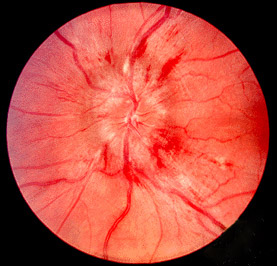
- Papilledema = blurred disc margins, loss of spontaneous venous pulsations — hallmark of chronic raised ICP (takes hours-to-days to develop, so may be absent acutely)
- CN VI (abducens) palsy = classic false-localizing sign (long intracranial course) causing horizontal diplopia
- Cushing triad (late): hypertension with a widened pulse pressure, bradycardia, irregular breathing
- Declining consciousness -> posturing (decorticate/flexor -> decerebrate/extensor) -> fixed pupils
- Numbers to know: normal ICP 5-15 mmHg; treat >20-22; CPP goal 60-70 mmHg
- ICP waveform: pathologic Lundberg A ("plateau") waves = sustained ICP elevations ~50 mmHg signaling exhausted compliance
Herniation Syndromes at a Glance
| Syndrome | What herniates | Classic findings | Vessel / complication |
|---|---|---|---|
| Subfalcine (cingulate) | Cingulate gyrus under the falx | Often silent; contralateral leg weakness | ACA compression |
| Uncal (transtentorial) | Uncus through tentorial notch | Ipsilateral blown pupil (CN III), "down-and-out" eye, contralateral hemiparesis | PCA infarct; Kernohan notch -> ipsilateral weakness (false localizing) |
| Central transtentorial | Diencephalon pushed downward | Progressive drop in consciousness, small -> fixed pupils, Cheyne-Stokes | Duret hemorrhages (midbrain/pons) |
| Tonsillar | Cerebellar tonsils through foramen magnum | Coma, apnea, cardiorespiratory collapse | Medullary compression -> death |
A 24-year-old man is admitted after a motorcycle crash with a temporal skull fracture. Over 2 hours his GCS falls from 14 to 7. The right pupil is fixed and dilated (7 mm), the right eye rests "down and out," and there is left-sided hemiparesis. BP 178/70, HR 46, breathing irregular.
- Diagnosis: Uncal (transtentorial) herniation. The uncus compresses the ipsilateral CN III — superficial parasympathetic fibers fail first, so a fixed dilated pupil precedes ptosis/ophthalmoplegia — and the cerebral peduncle, causing contralateral weakness. The likely lesion is an expanding epidural hematoma (biconvex/lens-shaped, middle meningeal artery, lucid interval).
- Next best step: Secure the airway; elevate head 30 degrees, give hyperosmolar therapy (mannitol or 3% saline) with brief hyperventilation to PaCO2 30-35 as a bridge, then emergent non-contrast CT and neurosurgical hematoma evacuation / decompression. Do not delay for prolonged workup.

Diagnosis & the Lumbar Puncture Trap
Non-contrast head CT is first-line — fast, and it detects hemorrhage, mass, midline shift, effaced sulci and basal cisterns, and hydrocephalus. MRI better characterizes tumor, edema, and early ischemia.
The board reflex: in a patient with papilledema, focal deficits, or a suspected mass lesion, do NOT perform a lumbar puncture before imaging — withdrawing CSF across a pressure gradient can precipitate tonsillar herniation. Get the CT first.
Definitive ICP monitoring is indicated in severe TBI (GCS <=8) with an abnormal CT. The external ventricular drain (EVD) is preferred because it both measures ICP and lets you drain CSF to treat it; an intraparenchymal "bolt" only measures. Fundoscopic papilledema and effaced basal cisterns on CT are key signs that intracranial compliance is spent.
A 29-year-old woman with obesity and recent isotretinoin use reports weeks of daily headache, pulsatile tinnitus, and transient visual obscurations. Exam shows bilateral papilledema and a CN VI palsy; the rest is normal. MRI/MRV is unremarkable (may show empty sella or flattened globes) — no mass, no venous thrombosis.
- Diagnosis: Idiopathic intracranial hypertension (pseudotumor cerebri) — classically an obese woman of childbearing age. Associations: vitamin A/retinoids, tetracyclines, growth hormone.
- Next best step: Image first to exclude a mass and cerebral venous sinus thrombosis (done here), then lumbar puncture — safe and diagnostic because pressure is diffuse and communicating with no mass: opening pressure >25 cm H2O with normal CSF composition. LP is also transiently therapeutic.
- Management: weight loss and acetazolamide (reduces CSF production); if vision is threatened, optic nerve sheath fenestration or a CSF shunt. Contrast this with the prior vignette: mass lesion = image first; IIH = LP is the answer.
Escalate in tiers:
- General: head of bed 30 degrees, head midline (aids venous drainage); avoid hypotension/hypoxia; treat fever, pain, and agitation; keep normoglycemia; avoid hypotonic fluids / hyponatremia
- Hyperosmolar therapy: mannitol 0.25-1 g/kg (watch for hypovolemia/renal injury) or hypertonic (3%) saline (preferred if hypotensive)
- Hyperventilation to PaCO2 ~30-35: rapid vasoconstriction, but a temporary bridge only — aggressive or prolonged use risks ischemia
- CSF drainage via EVD
- Refractory: deep sedation, barbiturate coma, decompressive craniectomy
- Treat the cause: evacuate hematoma; dexamethasone for vasogenic edema around tumor/abscess ONLY — steroids are not used in TBI (CRASH trial: increased mortality) or ischemic stroke
The Cushing triad signals impending herniation — a straining, slowing brainstem:
- Hypertension with a widened pulse pressure
- Bradycardia
- Irregular respirations (Cheyne-Stokes or ataxic)
Mechanism: as ICP rises, the reflex tries to force CPP = MAP - ICP back upward (sympathetic surge -> high BP -> baroreceptor-driven vagal bradycardia). On boards, the triad means act now — hyperosmolar therapy plus neurosurgery, not more workup. Beware the distractor of treating the hypertension: the BP is defending perfusion.
Practice Neuro now
Board-style questions, spaced-repetition flashcards, and a Socratic AI tutor — free to start.