Hypersensitivity Reactions (Types I–IV)
A high-yield Step 1 review of the four Gell-Coombs hypersensitivity types — IgE (I), antibody/cytotoxic (II), immune-complex (III), and delayed T-cell (IV) — with mechanisms, diagnostic tests, the anaphylaxis-vs-anaphylactoid distinction, and the classic vignette buzzwords boards reward.
The Gell-Coombs Framework
Hypersensitivity reactions are exaggerated or misdirected immune responses that damage the host. The classic Gell and Coombs classification splits them into four types by the effector mechanism, not the trigger. The single most testable organizing principle: Types I, II, and III are antibody (humoral) mediated, while Type IV is T-cell (cell) mediated — captured as "the first three are B, the fourth is T." Type I uses IgE; Types II and III use IgG/IgM; Type IV uses no antibody at all. Because the mechanisms overlap in real disease (e.g., hypersensitivity pneumonitis has both III and IV components), boards test the prototype disease for each type.
A C I D gives you Types I–IV in sequence:
- A — Anaphylactic / Atopic (Type I, IgE)
- C — Cytotoxic / antibody-mediated (Type II, IgG/IgM)
- I — Immune complex (Type III)
- D — Delayed / cell-mediated (Type IV, T cells)
Four Types at a Glance
| Type | Name | Mediator | Core mechanism | Classic examples |
|---|---|---|---|---|
| I | Immediate / anaphylactic | IgE on mast cells & basophils | Antigen cross-links preformed IgE → histamine, tryptase, leukotrienes (immediate min; late phase hrs) | Anaphylaxis, allergic rhinitis, asthma, atopic eczema, urticaria |
| II | Cytotoxic / antibody | IgG / IgM vs fixed cell-surface antigen | Opsonization/complement lysis, inflammation, or altered function | AIHA, ITP, Goodpasture, hemolytic disease of newborn, myasthenia gravis, Graves, pemphigus vulgaris |
| III | Immune complex | IgG immune complexes | Ag–Ab complexes deposit in tissue → complement → neutrophils | SLE, poststreptococcal GN, serum sickness, Arthus reaction, HBV-associated polyarteritis nodosa |
| IV | Delayed / cell-mediated | T cells (no antibody) | CD4⁺ → cytokines/macrophages; CD8⁺ → direct cytotoxicity; 48–72 h | Contact dermatitis (poison ivy, nickel), PPD/TB skin test, GVHD, acute graft rejection, granulomas |
- Types I–III are antibody-mediated; Type IV is T-cell mediated and is the only type transferable by T cells, not serum.
- Type I requires prior sensitization: first exposure sensitizes, re-exposure triggers. Symptoms in minutes (preformed histamine), late phase in hours (newly made leukotrienes + recruited eosinophils).
- Serum tryptase confirms mast-cell degranulation in anaphylaxis; first-line treatment of anaphylaxis is IM epinephrine (not antihistamines).
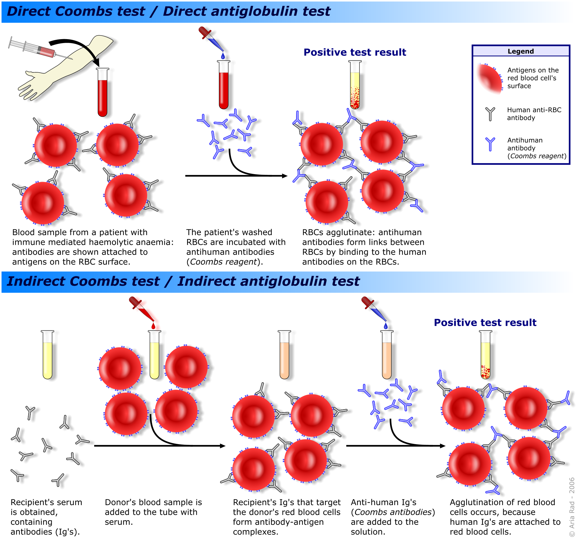
- Coombs test diagnoses Type II hemolysis: direct = antibody already on the patient's RBCs; indirect = antibody free in the patient's serum.
- Goodpasture = anti-GBM, LINEAR immunofluorescence (Type II); PSGN and SLE = GRANULAR / "lumpy-bumpy" immune-complex deposits (Type III).
- Antibody-mediated cellular dysfunction (myasthenia gravis anti-AChR, Graves anti-TSHR, pemphigus anti-desmoglein) is still classed as Type II, even though cells aren't destroyed.
- Transplant note: hyperacute rejection = Type II (preformed antibodies), acute cellular rejection = Type IV.

Match the buzzword to the type, then pick the next step:
- Minutes after a bee sting, a food (peanut/shellfish), or penicillin → hypotension, wheezing, angioedema, urticaria = Type I anaphylaxis → next best step: IM epinephrine; a rising tryptase confirms.
- The anaphylactoid mimic: radiocontrast dye, vancomycin (red-man syndrome), and opioids cause a clinically identical picture by direct mast-cell degranulation — non-IgE and needing no prior sensitization; treat it the same, but it is not true Type I hypersensitivity.
- ~5–10 days after a new drug (e.g., a β-lactam or antitoxin) → fever, urticarial rash, arthralgias, proteinuria, low C3/C4 = serum sickness (Type III).
- Hemoptysis + hematuria, linear IF on renal biopsy = Goodpasture (Type II).
- Young woman: malar rash, arthritis, proteinuria, low complement, anti-dsDNA = SLE (Type III).
- 48–72 h after gardening: linear vesicular rash on the forearms = poison-ivy contact dermatitis (Type IV); the same delayed timing explains induration at a PPD site.
Type II Sub-mechanisms & the Two Faces of Type III
Type II works three ways: (1) opsonization/complement lysis (autoimmune hemolytic anemia, transfusion reactions, hemolytic disease of the newborn); (2) complement- and Fc-driven inflammation recruiting neutrophils (Goodpasture, acute rheumatic fever); and (3) cellular dysfunction where antibody blocks or stimulates a receptor without killing the cell (myasthenia gravis, Graves, pemphigus vulgaris).
Type III has two named prototypes that are frequently confused. Serum sickness is systemic — circulating immune complexes form days after exposure to a foreign protein or drug and deposit in vessels, joints, and glomeruli. The Arthus reaction is local — intradermal antigen in someone with pre-existing IgG produces immune-complex deposition, edema, and necrosis at the injection site (classically after a vaccine booster).
Serum Sickness vs Arthus Reaction
| Feature | Serum sickness | Arthus reaction |
|---|---|---|
| Distribution | Systemic | Local (injection site) |
| Trigger | Foreign proteins / drugs acting as haptens | Intradermal antigen in a presensitized host |
| Antibody status | Antibodies form after exposure | Pre-existing circulating IgG |
| Onset | 5–10 days after exposure | Hours after injection |
| Findings | Fever, urticaria, arthralgias, lymphadenopathy, proteinuria, ↓C3/C4 | Local edema, erythema, induration, necrosis |
| Classic setting | Drug/antitoxin reaction | Vaccine booster (e.g., tetanus) |
Type IV (four) has 4 T's:
- T cells (CD4⁺ drive macrophages/granulomas; CD8⁺ kill directly)
- Transplant rejection (acute cellular)
- Tuberculin skin test (PPD) and other granulomatous disease
- Touching → contact dermatitis (poison ivy, nickel)
Remember it is delayed (48–72 h) and antibody-independent — the reason it's transferred by T cells, not serum.
Practice Immunology now
Board-style questions, spaced-repetition flashcards, and a Socratic AI tutor — free to start.