Hepatobiliary & Pancreatic Surgery
A Step 2 CK-focused walkthrough of hepatobiliary and pancreatic surgery, organizing gallstone complications, acute pancreatitis, and pancreatic cancer around ultrasound-first workup and next-best-step decisions. Emphasizes classic vignette buzzwords (Charcot/Reynolds, Courvoisier, double-duct sign) and when to choose HIDA, ERCP, or surgery.
The board framework: map the patient onto the obstruction spectrum
Hepatobiliary and pancreatic surgery on Step 2 CK is dominated by gallstone disease and its complications, plus acute pancreatitis and pancreatic cancer. The unifying skill is placing a patient on a spectrum of biliary obstruction caused by the same stones: transient cystic-duct obstruction (biliary colic), gallbladder inflammation (acute cholecystitis), common-bile-duct obstruction (choledocholithiasis), infected obstructed bile (ascending cholangitis), or pancreatic-duct obstruction (gallstone pancreatitis). Vignettes turn on three data points — fever/leukocytosis, the bilirubin/alk-phos pattern, and ultrasound (the first-line test for RUQ pain) — and on the single next best step. RUQ ultrasound is almost always the opening move; the branch points are when to add a HIDA scan, when to go to ERCP, and when to operate. Get those three decisions right and most questions in this block fall.
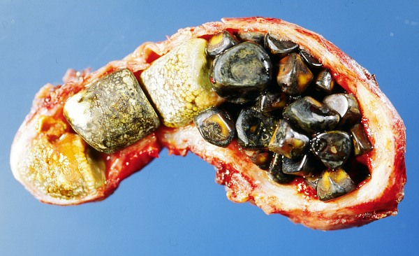
- Biliary colic: episodic RUQ/epigastric pain after fatty meals, resolves <6 h. No fever, normal WBC/LFTs. US shows stones → elective lap cholecystectomy.
- Acute cholecystitis: constant RUQ pain >6 h, fever, Murphy sign, leukocytosis; bili normal or mildly up. US: wall >3 mm, pericholecystic fluid, stones. Equivocal US → HIDA scan (gallbladder nonvisualization = positive). Tx: IV fluids, antibiotics, early lap chole, same admission.
- Choledocholithiasis: up direct bilirubin, up alk phos/GGT, dilated CBD >6 mm. Tx: ERCP extraction, then chole.
- Ascending cholangitis: Charcot triad (fever, jaundice, RUQ pain). Tx: fluids + antibiotics + urgent ERCP decompression.
- Gallstone pancreatitis: up lipase + stones on US.
- Acalculous cholecystitis: the ICU/TPN/burn/trauma patient → percutaneous cholecystostomy if too unstable for surgery.
- Porcelain gallbladder (calcified wall): risk factor for gallbladder cancer → cholecystectomy.
Distinguishing the four core biliary syndromes
| Condition | Pain | Fever/WBC | Bili / Alk phos | Imaging | Next step |
|---|---|---|---|---|---|
| Biliary colic | RUQ <6 h, postprandial | No | Normal | Stones, normal wall | Elective lap chole |
| Acute cholecystitis | RUQ >6 h, Murphy + | Yes | Normal / mild up | Wall >3 mm, pericholecystic fluid | Abx + early lap chole (HIDA if US equivocal) |
| Choledocholithiasis | RUQ, colicky | No / mild | up up direct bili, up alk phos | Dilated CBD >6 mm | ERCP, then chole |
| Ascending cholangitis | RUQ + jaundice | Yes (± shock/AMS) | up up bili + alk phos | Dilated CBD ± stone | Abx + urgent ERCP |
Vignette: A 68-year-old woman has 1 day of fever to 39°C, RUQ pain, and scleral icterus. BP 88/54, HR 112, and she is confused. WBC 18,000, total bilirubin 6.2, alk phos 410. US shows a dilated CBD with a stone.
Diagnosis: Ascending cholangitis. Fever + jaundice + RUQ pain = Charcot triad; adding hypotension and altered mental status = Reynolds pentad, signaling suppurative (obstructed, infected) cholangitis.
Next best step: Resuscitate with IV fluids and start broad-spectrum antibiotics, then proceed to urgent biliary decompression by ERCP (percutaneous transhepatic drainage or surgery if ERCP fails). Cholangitis is bile under pressure — antibiotics alone are not enough; decompression is definitive. Cholecystectomy follows once the patient stabilizes. Do not delay ERCP for a normal-appearing CBD if the clinical picture is cholangitis.
Vignette: A 45-year-old woman has severe epigastric pain radiating to the back with nausea and vomiting. Lipase 1,050 (>3× ULN). US shows gallstones; CBD not dilated. Triglycerides and calcium normal, minimal alcohol use.
Diagnosis: Acute gallstone pancreatitis. Diagnosis requires 2 of 3: characteristic pain, lipase/amylase >3× ULN, or imaging findings. Gallstones and alcohol are the two leading causes.
Next best step: Aggressive IV crystalloid, analgesia, and early enteral feeding as tolerated — prolonged NPO/TPN is outdated. Early CT is unnecessary and antibiotics are not given for uncomplicated pancreatitis. Add ERCP only for concurrent cholangitis or persistent biliary obstruction. For mild gallstone pancreatitis, perform cholecystectomy during the same admission once pain and enzymes improve, to prevent recurrence.
- Severity scoring: Ranson criteria, APACHE II, or BISAP. Ranson at admission (age >55, WBC >16k, glucose >200, LDH >350, AST >250) and at 48 h (Ca <8, Hct drop >10%, PaO2 <60, BUN rise >5, base deficit, fluid sequestration).
- Hemorrhagic signs: Cullen (periumbilical) and Grey Turner (flank) ecchymoses.
- Necrotizing pancreatitis: contrast CT shows nonenhancing pancreas. Infected necrosis (gas on CT or clinical deterioration) → antibiotics and a delayed step-up approach: percutaneous/endoscopic drainage first, then minimally invasive necrosectomy if needed, ideally after ~4 weeks. Routine CT-guided FNA is no longer required.
- Pseudocyst: encapsulated, amylase-rich fluid >4 weeks after onset, no epithelial lining. Observe if asymptomatic; drain via endoscopic cystogastrostomy if symptomatic, infected, or enlarging.
- Other: ARDS, hypocalcemia, splenic vein thrombosis → gastric varices, pseudoaneurysm.
Vignette: A 66-year-old smoker has 3 months of painless jaundice, dark urine, pale stools, and 8 kg weight loss. Exam reveals a palpable, nontender gallbladder (Courvoisier sign). Bilirubin 9, alk phos markedly elevated; CA 19-9 high.
Diagnosis: Pancreatic head adenocarcinoma obstructing the distal CBD. Migratory thrombophlebitis (Trousseau sign of malignancy) is a classic clue; CT may show the double-duct sign (dilated CBD and pancreatic duct).
Next best step: Pancreatic-protocol CT to stage and assess resectability (relationship to SMA/SMV/celiac axis). A resectable head tumor → pancreaticoduodenectomy (Whipple); most patients present with unresectable or metastatic disease and receive palliative biliary stenting plus chemotherapy. Board rule: painless jaundice + weight loss = pancreatic cancer until proven otherwise.
Charcot triad (ascending cholangitis) = Fever + Jaundice + RUQ pain.
Reynolds pentad = Charcot triad + hypotension + altered mental status → suppurative cholangitis needing urgent decompression.
Courvoisier sign = palpable nontender gallbladder + jaundice → suspect malignant distal CBD obstruction (pancreatic/biliary cancer), not stones (chronic stones scar the gallbladder, so it can't distend).
Causes of acute pancreatitis — I GET SMASHED: Idiopathic, Gallstones, Ethanol, Trauma, Steroids, Mumps, Autoimmune, Scorpion sting, Hypercalcemia/Hypertriglyceridemia, ERCP, Drugs.
Rigler triad (gallstone ileus) = pneumobilia + small-bowel obstruction + ectopic gallstone (typically lodges at the ileocecal valve); treat with enterolithotomy.

Practice Surgery now
Board-style questions, spaced-repetition flashcards, and a Socratic AI tutor — free to start.