Heart Failure: HFrEF vs HFpEF
Heart failure split by ejection fraction: HFrEF (systolic, EF ≤40%) versus HFpEF (diastolic, EF ≥50%) — their distinct patients, exam signs (S3 vs S4), workup, and why only HFrEF has the mortality-reducing "four pillars" of GDMT (with SGLT2 inhibitors now benefiting both).
The EF Split: Systolic vs Diastolic
Heart failure (HF) is a clinical syndrome in which the heart cannot deliver enough cardiac output to meet metabolic demand — or can do so only at the cost of elevated filling pressures. Boards divide HF by left ventricular ejection fraction (LVEF), measured on echocardiography:
- HFrEF (reduced, EF ≤40%) — a systolic problem: the ventricle cannot contract. The chamber dilates (eccentric hypertrophy) and stroke volume falls.
- HFpEF (preserved, EF ≥50%) — a diastolic problem: a stiff, thick ventricle cannot relax and fill. EF looks normal, but filling pressures are high.
- HFmrEF (mildly reduced, EF 41–49%) bridges the two.
Both share the same maladaptive neurohormonal cascade — RAAS and sympathetic activation — which initially compensates but ultimately drives remodeling, congestion, and progression. This is why the mortality-reducing drugs all block that cascade.
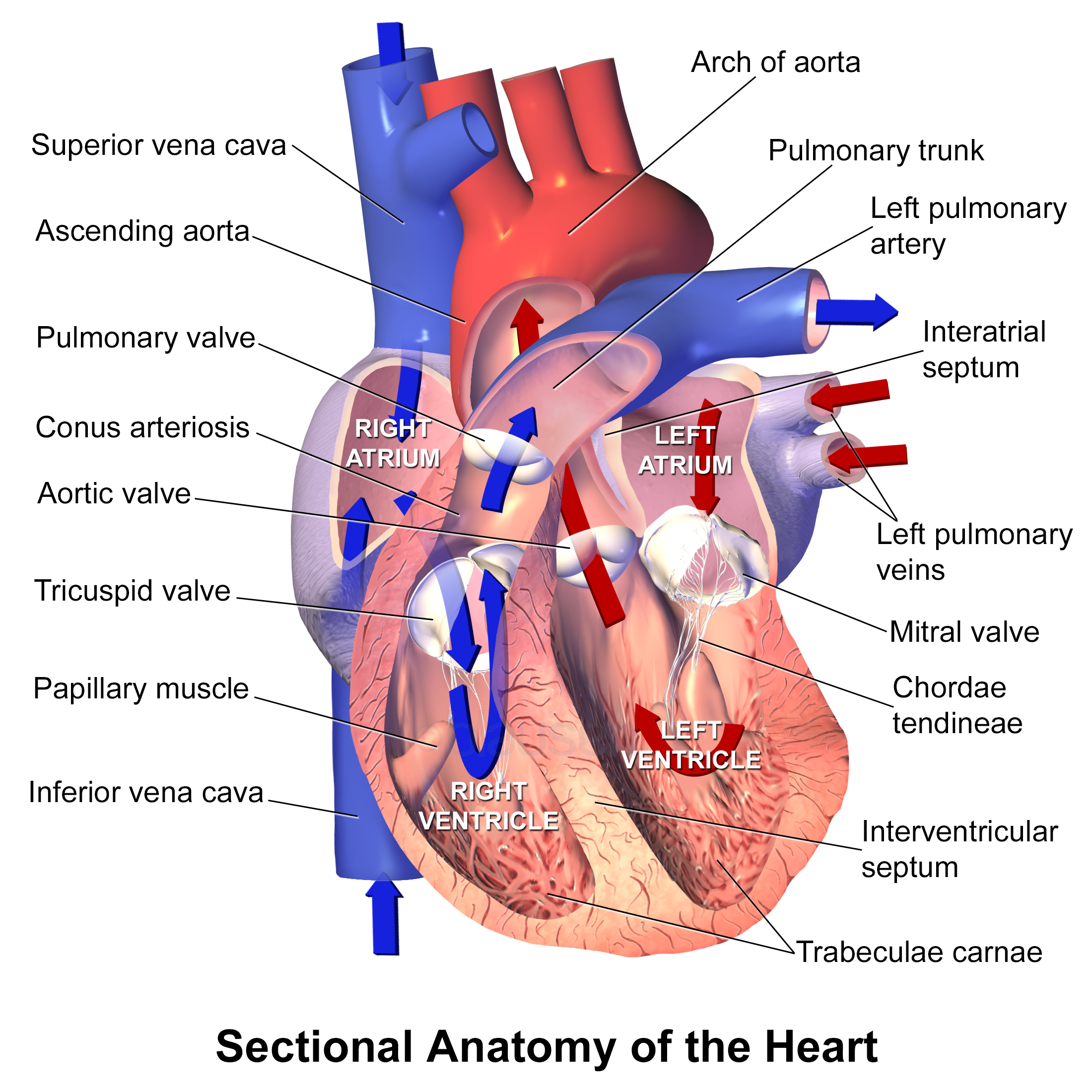
- HFrEF = EF ≤40% (systolic); HFpEF = EF ≥50% (diastolic); HFmrEF = 41–49%
- Transthoracic echocardiogram (TTE) is the key diagnostic test — it defines EF and separates the two types
- BNP / NT-proBNP rises in both; strong negative predictive value (good to rule out HF). Falsely low in obesity; falsely high in renal failure, AFib, elderly
- S3 gallop → volume overload / HFrEF; S4 gallop → stiff ventricle / HFpEF
- Classic HFrEF cause = ischemia / prior MI; classic HFpEF cause = hypertension (think elderly, obese, diabetic woman)
- Only HFrEF has robust mortality-reducing GDMT; HFpEF long had none — now SGLT2 inhibitors benefit both
- Loop diuretics relieve congestion but do NOT improve survival in either type
HFrEF vs HFpEF
| Feature | HFrEF | HFpEF |
|---|---|---|
| Ejection fraction | ≤40% | ≥50% |
| Core defect | Systolic (contraction) | Diastolic (relaxation/filling) |
| Ventricular geometry | Dilated, eccentric hypertrophy | Concentric hypertrophy, small stiff cavity |
| Classic patient | Post-MI, dilated cardiomyopathy, younger | Elderly, hypertensive, obese, diabetic, female |
| Classic cause | Ischemia / CAD | Hypertension |
| Extra heart sound | S3 | S4 |
| Mortality-reducing Rx | Four pillars of GDMT | SGLT2 inhibitor (rest treats symptoms/comorbidities) |
Treatment: The Four Pillars
Foundational therapy for HFrEF is the "four pillars" of guideline-directed medical therapy (GDMT), each independently proven to reduce mortality:
- ARNI (sacubitril–valsartan) — preferred over an ACEi/ARB
- Beta-blocker — only carvedilol, metoprolol succinate, or bisoprolol have mortality data; start when euvolemic, never during acute decompensation
- MRA — spironolactone or eplerenone
- SGLT2 inhibitor — dapagliflozin or empagliflozin
Add hydralazine + isosorbide dinitrate in self-identified Black patients or those intolerant of ACEi/ARB. Loop diuretics (furosemide) treat congestion only. Devices: ICD for primary prevention of sudden death if EF ≤35% despite ≥3 months of GDMT; CRT if EF ≤35% + LBBB + QRS ≥150 ms.
For HFpEF: control blood pressure, rate-control AFib, relieve volume with diuretics, treat comorbidities — and add an SGLT2 inhibitor, the first class with clear benefit here.
- HFrEF vignette: a man months after an anterior MI with progressive dyspnea, a laterally displaced PMI, an S3 gallop, and echo showing EF 30% → start the four pillars once stable.
- HFpEF vignette: an elderly obese hypertensive woman with exertional dyspnea, an S4 gallop, bibasilar crackles, and echo showing normal EF with LVH / diastolic dysfunction.
- Suspected new HF — next best step: check BNP/NT-proBNP and obtain a transthoracic echo to define EF.
- Acute decompensated HF: orthopnea, PND, crackles, JVD, Kerley B lines and cephalization on CXR → next best step = IV loop diuretic (plus O2; nitrates if hypertensive).
- Trap: do not start a beta-blocker in an acutely volume-overloaded patient — wait until euvolemic. Beta-blockers are for chronic stable HFrEF.
LMNOP — the classic bundle for acute decompensated heart failure / flash pulmonary edema:
- L — Lasix (IV loop diuretic — the mainstay)
- M — Morphine (↓ preload/anxiety; used less now)
- N — Nitrates (venodilation → ↓ preload)
- O — Oxygen
- P — Position upright / Positive-pressure ventilation (CPAP/BiPAP)
Left vs Right Heart Failure
| Feature | Left HF | Right HF |
|---|---|---|
| Congestion backs up into | Lungs (pulmonary) | Systemic veins |
| Symptoms / signs | Dyspnea, orthopnea, PND, crackles, pulmonary edema | JVD, peripheral edema, hepatomegaly, ascites |
| Classic clue | Kerley B lines, pink frothy sputum | Hepatojugular reflux, nutmeg liver |
| Most common cause | Ischemia, hypertension | Left heart failure (then cor pulmonale) |

Practice Cardiology now
Board-style questions, spaced-repetition flashcards, and a Socratic AI tutor — free to start.