Glaucoma
A boards-focused glaucoma lesson contrasting chronic primary open-angle disease (painless peripheral loss, cupping, latanoprost first-line) with acute angle-closure glaucoma (a painful red-eye emergency treated by rapid IOP lowering and definitive laser peripheral iridotomy), including next-best-step vignettes, a comparison table, and drug-mechanism mnemonics.
What glaucoma is
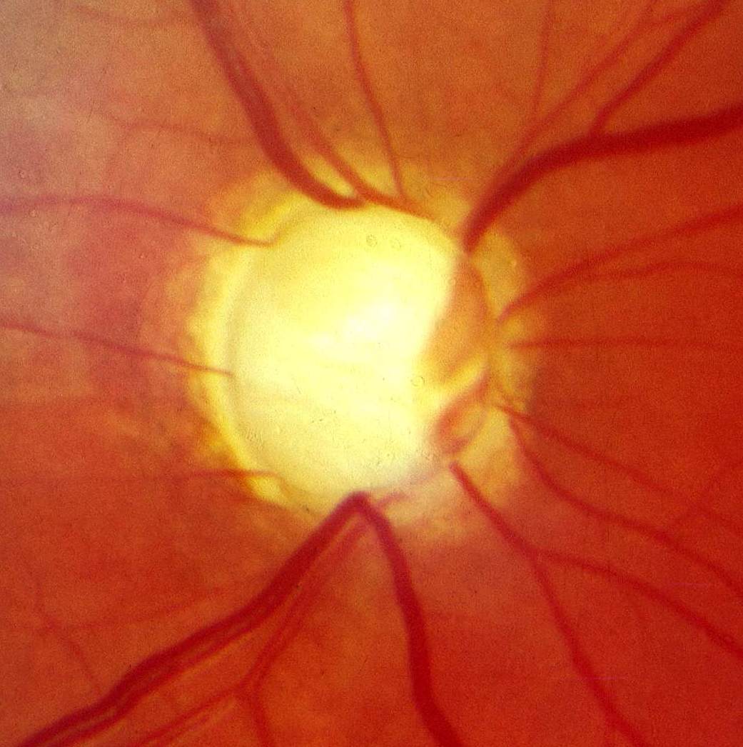
Glaucoma is a progressive optic neuropathy defined by characteristic cupping of the optic disc (↑ cup-to-disc ratio) and matching visual field loss. Elevated intraocular pressure (IOP) is the major modifiable risk factor but is not required for the diagnosis — normal-tension glaucoma occurs with IOP ≤21 mmHg. Aqueous humor is produced by the ciliary body and drains via the trabecular meshwork → canal of Schlemm (conventional, the majority of outflow) plus the uveoscleral route; impaired outflow raises IOP and damages retinal ganglion cell axons. Boards divide glaucoma into primary open-angle (chronic, painless, most common) and angle-closure (which can present as an acute, sight-threatening emergency). The two highest-yield skills are recognizing the silent peripheral vision loss of open-angle disease and treating acute angle-closure immediately.
- Most common glaucoma in the US; painless, bilateral, gradual
- Risk: older age, African ancestry, positive family history, ↑ IOP, thin central cornea, myopia (diabetes is a weaker/debated association)
- Mechanism: ↓ aqueous outflow through the trabecular meshwork; the angle is OPEN on gonioscopy
- IOP is often >21 mmHg but may be normal (normal-tension glaucoma)
- Signs: ↑ cup-to-disc ratio (>0.5 or asymmetric), arcuate (Bjerrum) scotomas, nasal step; peripheral loss progresses to tunnel vision with central acuity spared until late
- "Silent thief of sight" — asymptomatic early, usually caught on screening
- Workup: tonometry (IOP), dilated fundoscopy (disc cupping), automated perimetry (visual fields), gonioscopy (open angle), OCT of the retinal nerve fiber layer
- First-line for POAG: topical prostaglandin analog (latanoprost) — ↑ uveoscleral outflow, once nightly; classic side effects: eyelash lengthening, iris/periocular hyperpigmentation, conjunctival hyperemia
- ↓ aqueous production: beta-blocker (timolol), alpha-2 agonist (brimonidine), carbonic anhydrase inhibitor (topical dorzolamide; systemic acetazolamide)
- ↑ aqueous outflow: prostaglandins (uveoscleral) and pilocarpine/cholinergics (ciliary-muscle contraction → opens the trabecular meshwork; miosis opens a closed angle)
- Escalation: laser trabeculoplasty (SLT — now also an accepted first-line option) → incisional surgery (trabeculectomy, tube shunt)
- Timolol caution: avoid topical beta-blockers in asthma/COPD, bradycardia, or heart block
- Goal of therapy is slowing progression by lowering IOP; existing field loss is not reversible
Vignette: A 63-year-old African American man has a routine eye exam. He is asymptomatic, but IOP is 27 mmHg bilaterally and the cup-to-disc ratio is 0.7. On questioning, he admits to occasionally "missing" cars merging from the side while driving.
Diagnosis: Primary open-angle glaucoma.
Next best step: Automated perimetry (visual field testing) to document characteristic defects (arcuate scotoma / nasal step) and establish a baseline; gonioscopy confirms an open angle.
Treatment: Begin a topical prostaglandin analog (latanoprost) and monitor IOP and fields over time.
Vignette: A 58-year-old hyperopic woman develops sudden severe right-eye pain, frontal headache, nausea, and vomiting, and sees halos around lights after sitting in a dark theater (or after starting an anticholinergic). Exam: red eye, a mid-dilated, fixed, poorly reactive pupil, a hazy/'steamy' cornea, and a rock-hard globe.
Diagnosis: Acute angle-closure glaucoma (pupillary block).
Next best step: Tonometry confirms markedly elevated IOP (often >40 mmHg) → emergent IOP-lowering: topical timolol + apraclonidine/brimonidine + pilocarpine, plus systemic acetazolamide (± IV mannitol). Note: pilocarpine may not work until IOP is lowered, because the high-pressure iris sphincter is ischemic.
Definitive treatment: laser peripheral iridotomy. Avoid mydriatics/atropine — dilation worsens the block.

Open-angle vs. acute angle-closure
| Feature | Open-angle (POAG) | Acute angle-closure |
|---|---|---|
| Onset | Gradual, chronic | Sudden — emergency |
| Pain | Painless | Severe pain, headache, N/V |
| Vision | Slow peripheral loss → tunnel | Blurred, halos around lights |
| Pupil | Normal | Mid-dilated, fixed |
| Cornea | Clear | Hazy / steamy |
| IOP | Often >21 (may be normal) | Markedly ↑ (often >40) |
| Gonioscopy | Open angle | Closed angle |
| Key risk factors | African ancestry, myopia, thin cornea | Hyperopia, Asian ancestry, shallow AC, dim light |
| Definitive treatment | Lower IOP (drops → laser/surgery) | Laser peripheral iridotomy |
Organize glaucoma drugs by mechanism:
- Decrease aqueous PRODUCTION → "BAC": Beta-blocker (timolol), Alpha-2 agonist (brimonidine), Carbonic anhydrase inhibitor (dorzolamide / acetazolamide)
- Increase OUTFLOW: Prostaglandins (uveoscleral) and Pilocarpine (cholinergic — ciliary-muscle contraction opens the trabecular meshwork; miosis opens a closed angle)
Latanoprost buzzwords: Lashes lengthen and the iris darkens (hyperpigmentation) — the classic tested side effects.
Angle-closure rule: the pinpoint miotic (pilocarpine) opens the angle, whereas mydriatics/anticholinergics close it — never pharmacologically dilate an eye with suspected angle-closure.
Practice Ophthalmology now
Board-style questions, spaced-repetition flashcards, and a Socratic AI tutor — free to start.