GI Tract Histology
A board-focused walk through GI tract histology: the universal four-layer wall plan and enteric plexuses, the epithelial transitions and cell types of each segment, and the classic disease correlations (Barrett metaplasia, Hirschsprung aganglionosis, pernicious anemia).
The one blueprint, repeated
From esophagus to anus the gut is built on one repeating four-layer plan: mucosa (epithelium + lamina propria + muscularis mucosae) → submucosa → muscularis externa (inner circular + outer longitudinal smooth muscle) → serosa/adventitia. What changes down the tube is the epithelium and the regional add-ons layered onto that plan. Boards hammer two themes. First, the abrupt epithelial transitions — stratified squamous esophagus → simple columnar stomach (Z-line), and simple columnar rectum → stratified squamous anus (pectinate line) — because these are exactly where metaplasia and cancer arise. Second, the enteric nervous system: the submucosal (Meissner) and myenteric (Auerbach) plexuses, whose loss defines Hirschsprung disease. Learn the normal cell types and most diseases fall out as either loss of a cell (parietal → pernicious anemia) or metaplasia of an epithelium (squamous → columnar in Barrett).
- Mucosa = epithelium + lamina propria (loose CT, immune cells) + muscularis mucosae (thin smooth muscle)
- Submucosa = dense CT + submucosal (Meissner) plexus; holds the glands in duodenum (Brunner) and esophagus
- Muscularis externa = inner circular + outer longitudinal, with the myenteric (Auerbach) plexus sandwiched between
- Serosa (mesothelium) covers intraperitoneal gut; adventitia anchors retroperitoneal + thoracic esophagus
- Meissner → Secretions & blood flow; Auerbach → Motility
- Surface-area amplifiers (small bowel): plicae circulares > villi > microvilli
- Esophagus muscle: upper 1/3 skeletal, middle mixed, lower 1/3 smooth
- Hirschsprung = congenital absence of BOTH plexuses (failed neural crest migration) → aganglionic, tonically contracted distal colon

Segment-by-segment histology
| Segment | Epithelium | Board-tested hallmark |
|---|---|---|
| Esophagus | Nonkeratinized stratified squamous | Skeletal→smooth muscle down the tube; submucosal mucous glands |
| Stomach | Simple columnar | Gastric pits, rugae; parietal + chief cells |
| Duodenum | Simple columnar + villi | Brunner glands in submucosa (alkaline mucus) |
| Jejunum | Simple columnar + villi | Tallest plicae circulares, long villi |
| Ileum | Simple columnar + villi | Peyer patches (M cells); goblet cells more numerous than proximal small bowel |
| Colon | Simple columnar, no villi | Crypts packed with goblet cells (densest in gut); teniae coli |
| Anus | Stratified squamous (distal) | Pectinate line transition |
Gastric mucosal cells
| Cell | Secretes | Location | Board link |
|---|---|---|---|
| Parietal (oxyntic) | HCl + intrinsic factor | Body/fundus | Autoimmune loss → pernicious anemia (B12) |
| Chief (zymogenic) | Pepsinogen (+ gastric lipase) | Gland base | Basophilic, RER-rich |
| Mucous neck/surface | Mucus + bicarbonate | Neck/surface | Protective barrier |
| G cell | Gastrin | Antrum | Gastrinoma → Zollinger-Ellison |
| ECL cell | Histamine | Body/fundus | Drives parietal acid; gastric carcinoid |
| D cell | Somatostatin | Antrum/body | Inhibits gastrin + acid |
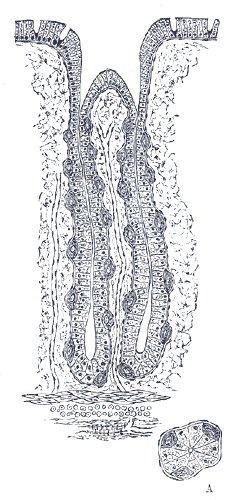
- Brunner glands = submucosal, alkaline (bicarb-rich) mucus; duodenum ONLY; hypertrophy with peptic ulcer disease
- Paneth cells = base of the crypts of Lieberkühn; eosinophilic granules of lysozyme, defensins, TNF-α (antimicrobial)
- Peyer patches = aggregated lymphoid follicles in lamina propria/submucosa of the ileum; M cells sample luminal antigen
- Goblet cells increase distally: fewest in duodenum → densest in the colon
- Villi = small bowel only; absent in colon (flat surface, deep crypts)
- Crypts house stem cells that regenerate the epithelium every ~3–5 days
- Celiac disease → villous blunting, crypt hyperplasia, intraepithelial lymphocytes, worst in duodenum/proximal jejunum
Stem: A 55-year-old man with 10 years of heartburn undergoes EGD. Distal esophageal biopsy shows columnar epithelium with goblet cells replacing the normal squamous lining.
Diagnosis: Barrett esophagus — intestinal metaplasia from chronic GERD acid injury (nonkeratinized stratified squamous → simple columnar with goblet/mucous cells).
Why it matters: metaplasia → dysplasia → esophageal adenocarcinoma (distal 1/3).
Next best step: endoscopic surveillance biopsies + PPI therapy; for high-grade dysplasia, endoscopic resection/ablation.
Stem: A neonate fails to pass meconium within 48 hours, then develops bilious vomiting and abdominal distension. Contrast enema shows a narrow distal segment with proximal dilation.
Diagnosis: Hirschsprung disease — failed neural crest migration → absent ganglion cells in the submucosal (Meissner) AND myenteric (Auerbach) plexuses of the distal colon/rectum; the aganglionic segment stays contracted. Associated with Down syndrome.
Confirm (next best step): rectal suction biopsy = gold standard (shows absent ganglia).
Management: surgical resection of the aganglionic segment (pull-through).
Plexuses (letters match):
- Submucosal / Meissner → Secretions & blood flow
- Myenteric / Auerbach → Motility
Gastric cells:
- PARIETAL cells Pump Protons (HCl) + make intrinsic Factor
- CHIEF cell = the chef → cooks pepsinogen
- G cell → Gastrin
Location: Brunner glands sit Below the mucosa (submucosa) in the duodenum; Peyer patches Populate the ileum.
Practice Histology now
Board-style questions, spaced-repetition flashcards, and a Socratic AI tutor — free to start.